

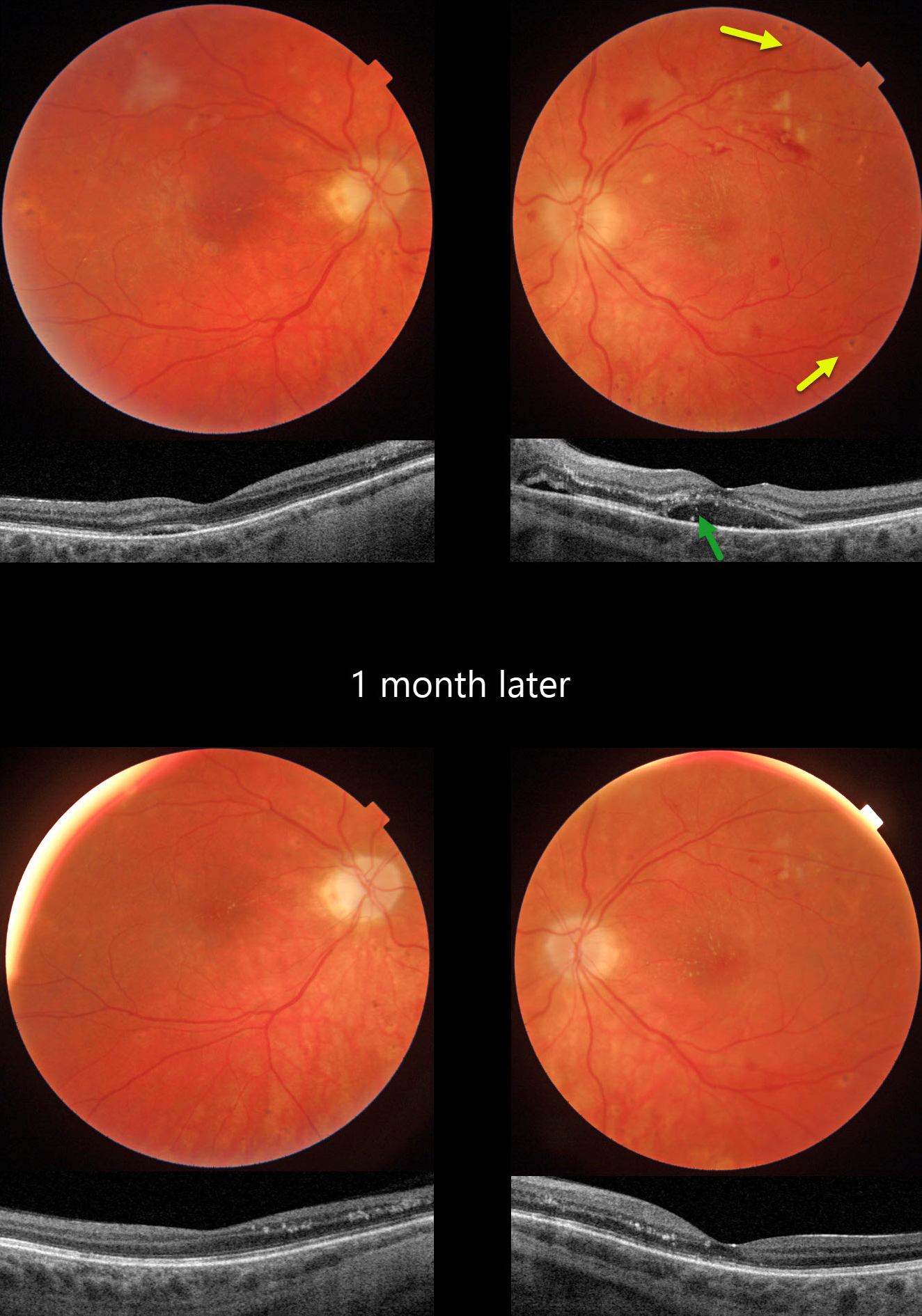
This 21YO female presented with bilateral vision loss. She was admitted to the hospital 2 weeks earlier for malignant hypertension (HTN), likely from methamphetamine abuse. Blood pressure at that time was 292/208. Vision was hand motion OD and 20/80 OS.
Triton color photography of her left eye shows a somewhat pale and swollen nerve, mostly inner retinal hemorrhages, and resolving nerve fiber layer infarcts (cotton-wool spots, CWS). Faint lipid exudates radiate nasally and superiorly from the macular center. Multifocal small hyperpigmented choroidal lesions are noted outside the arcades. Swept-source OCT shows subretinal fluid with hyperreflective lipid in the subretinal space and outer nuclear layer. Similar but much milder findings are noted in her right eye.
When examined 1 month later, vision remained at hand motion OD and improved to 20/40 OS with improved funduscopic and OCT findings.
Learning Points:
Malignant hypertension, defined as blood pressure above 180 systolic and/or 120 diastolic, is a life-threatening medical emergency. Eye doctors are in a unique position to often diagnose this condition. Patients will often present with bilateral optic nerve swelling, flame-shaped retinal hemorrhages, CWS, and, with more chronic disease, lipid precipitates in the nasal macular outer plexiform layer.
Acute hypertensive choroidopathy outside the setting of pre-eclampsia, in our experience, is quite rare. Independent of the disc and neurosensory retinal findings, patients present with localized multifocal serous retinal detachments (de Venecia and Jampol, Arch Ophthalmol 1984;102:68-73). With blood pressure control, these resolve often with minimal funduscopic changes. Focal (Elschnig spots) or linear (Siegrist streaks) choroidal pigmentary changes may result from more severe disease.



