

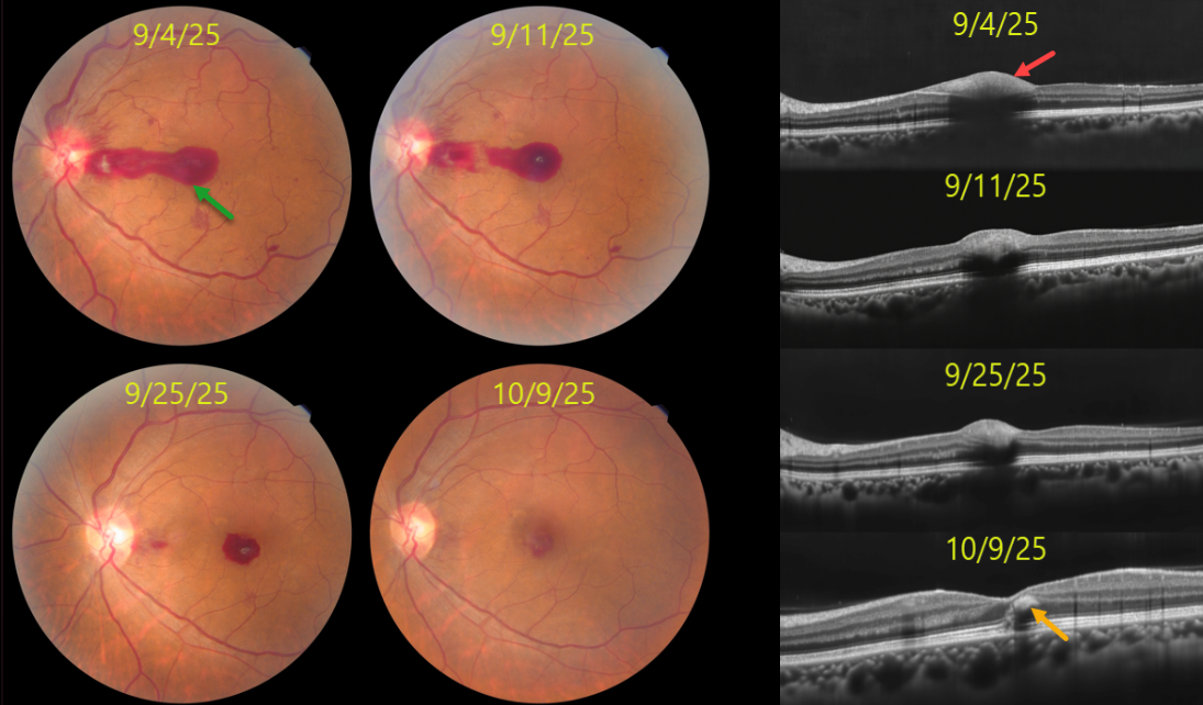
This 44YO female presented with sudden vision loss OS with uncontrolled hypertension (180/110 mmHg). Vision was 20/20 OD and 20/200 OS.
Color photography shows a pre-retinal hemorrhage extending from the temporal edge of the disc through the central macula, disc and peripapillary inner retinal hemorrhages, and blot and nerve fiber layer hemorrhages. The pre-retinal blood localizes to the sub-internal limiting membrane (ILM) space on OCT. The retinal findings normalized over the next 4-5 weeks with visual improvement to 20/20. OCT at the final visit shows a small amount of mid-retinal foveal blood.
Learning Points:
Malignant hypertension, defined as blood pressure above 180 systolic and/or 120 diastolic, is a life-threatening medical emergency. Eye doctors are often uniquely positioned to diagnose this condition (Shukla et al, Indian J Ophthalmol 2025;73:1552-1555). Patients will often present with bilateral optic nerve swelling, flame-shaped retinal hemorrhages, CWS, and, with more chronic disease, lipid precipitates in the nasal macular outer plexiform layer.
Sub-ILM hemorrhage, to the best of our knowledge, has not been previously described in hypertensive retinopathy. It is usually associated with retinal arterial macroaneurysm, Valsalva retinopathy, trauma, and blood dyscrasias. In our patient’s case, we hypothesize that the sub-ILM likely originated from the disc and dissected temporally across the macula.



