

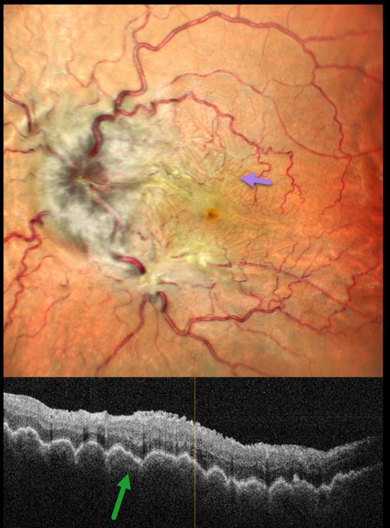
This 44YO male presented with 2 months of decreased vision in his left eye. A retinal detachment repair was done 9 months earlier with vitrectomy and silicone oil, followed by oil removal 3 months later. Vision was 20/20 in his normal OD and 20/90 OS. The intraocular pressure (IOP) was 10 mmHg OD and 3 mmHg OS. Pseudophakodonesis of the IOL was present OS.
Pseudocolor SLO imaging shows a moderately swollen nerve, tortuous retinal vessels, and extensive, irregularly oriented chorioretinal folds throughout the posterior pole. OCT shows a thickened choroid with overlying undulating chorioretinal folds.
We advised IOL explantation with vitrectomy and silicone oil, but he was immediately lost to follow-up.
Learning Points:
Hypotony maculopathy most commonly develops following surgery (including glaucoma filtration surgery and wound leaks) and trauma. Findings include low IOP, chorioretinal folds, and occasionally optic disc edema. Findings usually promptly resolve once the IOP normalizes.



