

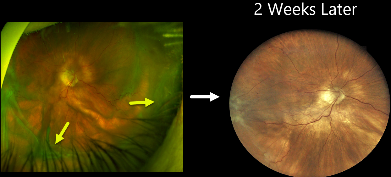
This 84YO female received an intravitreal Eylea HD injection in her right eye on 12/28/23 for macular edema from a branch retinal vein occlusion. She presented on 1/18/24, complaining of waking several days earlier with severe painless vision loss in this eye. She denied trauma or Valsalva. Vision was counting fingers, and intraocular pressure was 1 mmHg. There were no apparent wound leaks or conjunctival blebs.
Optos color RG imaging shows severe, diffuse, irregular scleral folds without choroidal detachment.
Treatment was initially conservative with topical atropine, prednisolone acetate, and oral steroids. However, findings were stable, and IOP remained at 1 mmHg after 3 days. The patient then underwent ultrasound biomicroscopy (UBM) to assess for a suspected posterior cyclodialysis cleft, followed by cryotherapy.
One week later, we planned on injecting an expandable C3F8 gas bubble in hopes of normalizing the eye pressure. When she presented on 2/1/24 for this procedure, her vision remained at counting fingers, but her IOP had increased to 2 mmHg. Fundus examination was nearly normal with almost complete resolution of the scleral folds.
Learning Points:
Although the definitive etiology of our patient’s hypotony remains unclear, our working theory is either a posterior cyclodialysis cleft or a very slow-leaking injection wound. As described by Lee et al. (Am J Ophthalmol Case Rep. 2021 Sep; 23: 101134), incomplete penetration into the vitreous cavity, early injection, or continued injection on needle withdrawal could lead to the formation of a posterior cyclodialysis cleft during intravitreal injections. No cleft was visible on gonioscopy, but a strong suspicion of a posterior cleft was raised on UBM. Our other differential included a slow-leaking scleral wound from a prior injection. No leak was visible at initial examination, but with an IOP of 1 mmHg, a spontaneous leak would not be expected. Both these theories are confounded by a history of severe (-15D) myopia prior to cataract surgery, with possible thinned sclera predisposing her to the hypotonus scleral folds.



