

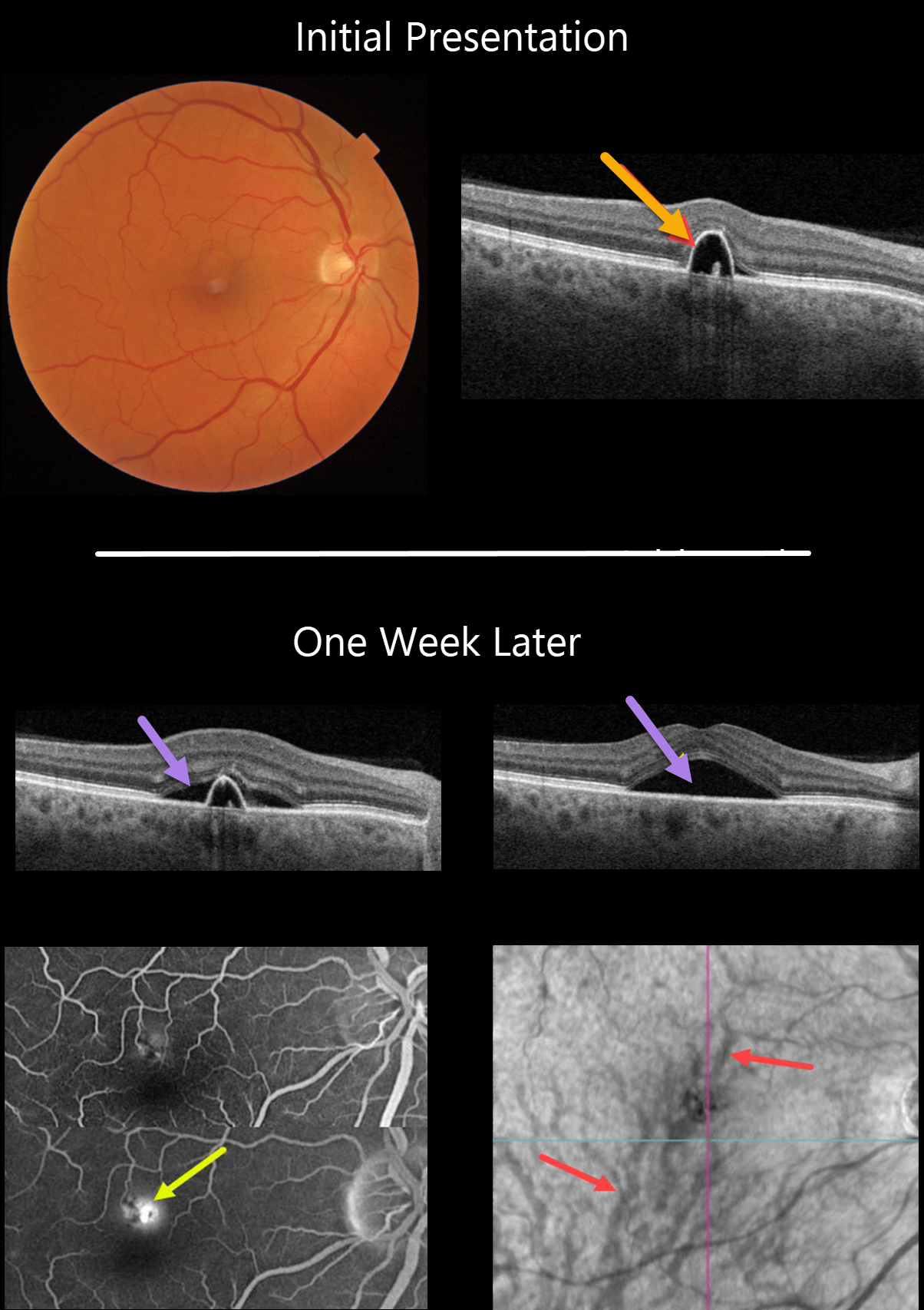
This 55YO male presented on 1/24/22 with a small symptomatic pigment epithelial detachment (PED) in the superior right fovea. Vision was 20/30.
One week later, he was subjectively worse, although vision remained at 20/30. There was a new small blister of foveal subretinal fluid, best captured on swept-source OCT.
Optos fluorescein angiography shows a typical blot leak along the dome of the PED. En face OCT shows a choroidal venous anastomosis.
Learning Points:
The choroidal venous system, as with the neurosensory retinal vasculature, usually respects the horizontal meridian. With pachychoroid disorders, these anastomoses develop, and in this patient’s case, the inferior vortex system drains nearly the entire macula.
For a wonderful review highlighting the concept of venous overload choroidopathy, see Spaide et al, Progress Retinal Eye Research 2022;86;100973.



