

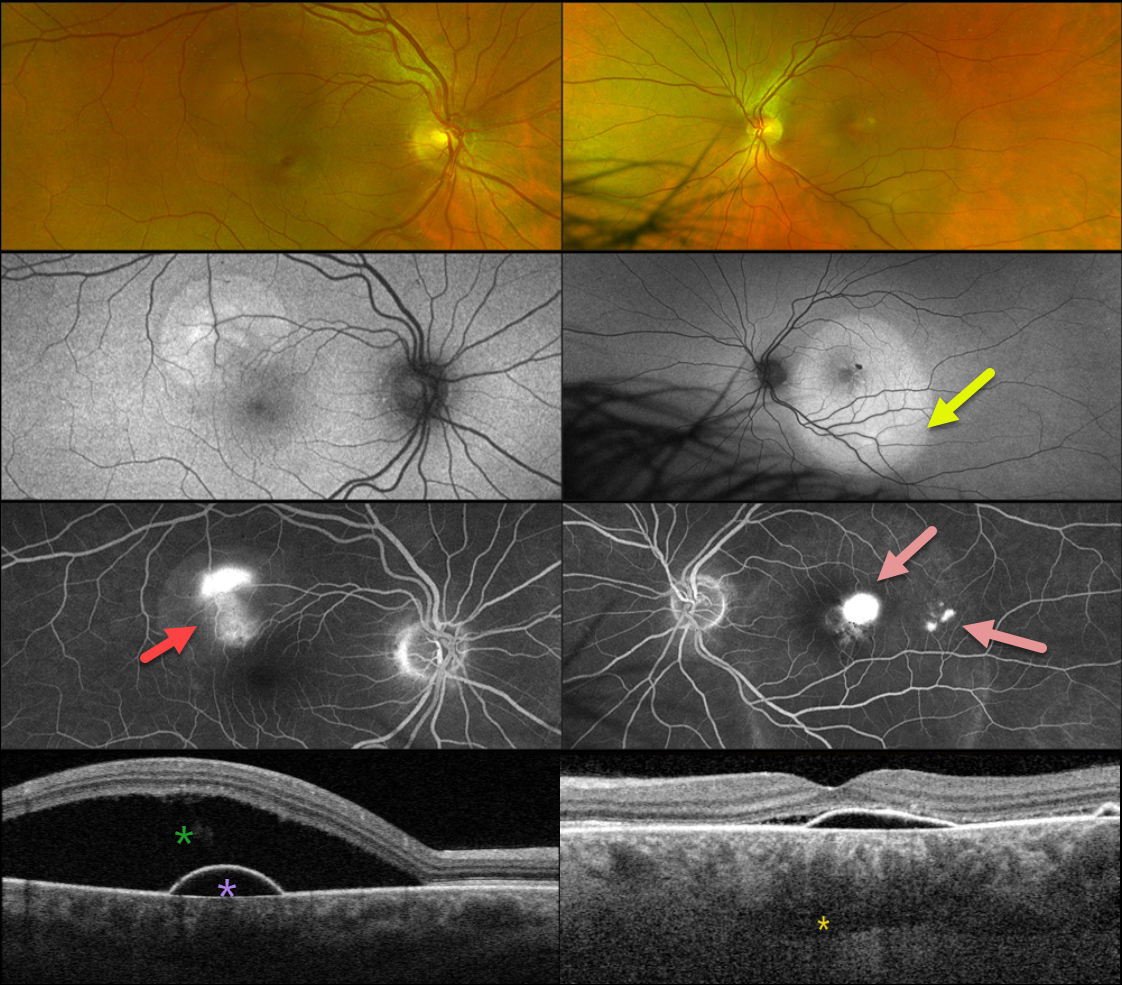
This healthy 48YO male presented with 1-2 weeks of decreased vision. There was no past ocular history. Vision was 20/25 OD and 20/70 OS.
Optos color RG imaging shows a superotemporal serous detachment OD with a larger serous detachment throughout the left macula. Triton swept-source OCT shows a shallow RPE detachment with some nasal subretinal fluid. Loculated outer choroidal fluid is seen. A more dramatic serous detachment with an underlying RPE detachment is noted OD.
On fundus autofluorescence, these serous detachments show hyper-FAF. Fluorescein angiography shows a smokestack leak OD with multifocal blot leaks OS.
We will initially observe our patient and recommend photodynamic therapy if the fluid fails to resolve over the coming months.
Learning Points:
Initially described by Spaide and Ryan (AJO 2015;160:1211-1216), loculated outer choroidal fluid is found in about two-thirds of patients with idiopathic central serous chorioretinopathy (ICSC). It is best appreciated with swept-source or enhanced depth imaging OCT.
This fluid most commonly interdigitates with the larger outer choroidal vessels, as in our patient, but can rarely appear in the suprachoroidal space. It is more common with a thicker choroid and sclera (AJO 2022;235:197-203).



