

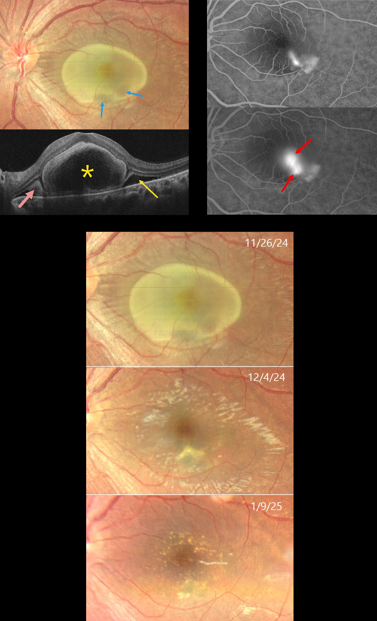
This 31YO female complained of decreased vision in her left eye for 6 weeks. She was diagnosed elsewhere with cysticercosis and referred to us for further evaluation. Vision was 20/20 in her normal OD and 20/200 OS.
Color photography shows a yellow oval lesion occupying nearly the entire macula. OCT scanning shows a large subfoveal bacillary layer detachment (BALAD). Hyperreflective fibrin likely lines the inner border of the BALAD. Subretinal fibrin and subretinal fluid (SRF) are noted on either edge of the BALAD. Fluorescein angiography shows either a smokestack or two independent blot leaks with staining inferotemporally. The yellow foveal lesion is relatively depigmented within the region of the angiographic leakage. The lesion and fluid spontaneously resolved over about 6 weeks, with residual drusen-like changes. Vision at the final visit was 20/20.
Learning Points:
In idiopathic central serous chorioretinopathy (ICSC), fluorescein dye diffuses through a physiological break in the RPE and then pools within the subretinal space. The clearing within our patient’s inferotemporal subretinal fibrin (as evidenced by the hyporeflective temporal SRF on OCT) is likely caused by serous fluid entering the subretinal space and ‘washing away’ some of the fibrin.
Most patients with ICSC have a single, unilateral leak. Blot leaks are most common, with only 10% of cases exhibiting a smokestack. The multifocal leaks and subretinal fibrin noted in our patient are more commonly found in steroid-induced disease, although our patient denied steroid exposure. Bacillary layer (cone and rod inner and outer segments) detachment appears as a unique dome-shaped collection of intraretinal fluid from photoreceptor splitting. These detachments can be observed in an increasing number of conditions, most classically Vogt-Koyanagi-Harada disease (Cicinelli et al, Ophthalmology Retina 2020;4:454-456). BALAD is also a biomarker for severe ICSC with steroid use (Casella et al, Int J Retina Vitreous 2024;10;96).



