

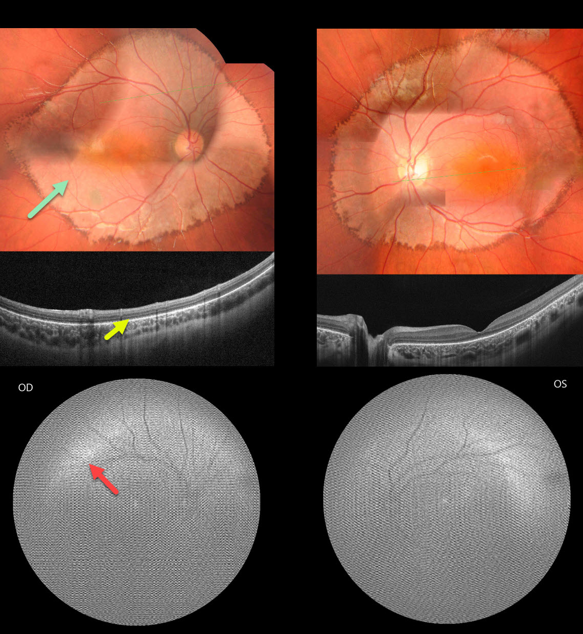
This healthy 6YO boy presented with diminished vision noticed by his parents and teacher. There was no family history of eye disease. Vision was 20/200 OU.
Fundus photography shows fairly symmetrical, creamy, pale subretinal lesions encircling each posterior pole. The margins of these lesions have a somewhat irregular hyperpigmented border.
OCT shows outer retinal thinning and loss of the outer retinal band detail, along with a thinned or absent RPE band.
Fundus autofluorescence (FAF) was of extremely poor quality, and possibly shows superior and temporal hyper-FAF just outside each macula. It was quite difficult to obtain imaging in our patient, and these studies required multiple sessions.
Learning Points:
Unfortunately, we do not have a specific diagnosis for our patient’s presumed sporadic inherited retinal disease (IRD). The location and bilaterality of the multimodal findings suggest a cone-mediated disorder. Our patient’s funduscopic appearance somewhat resembles that found in autosomal recessive CDH3 disease, which includes sparse scalp hair and occasional limb abnormalities (Hull et al, JAMA Ophthalmol 2916;134:992-1000). Genetic testing could not be performed due to cost constraints.



