

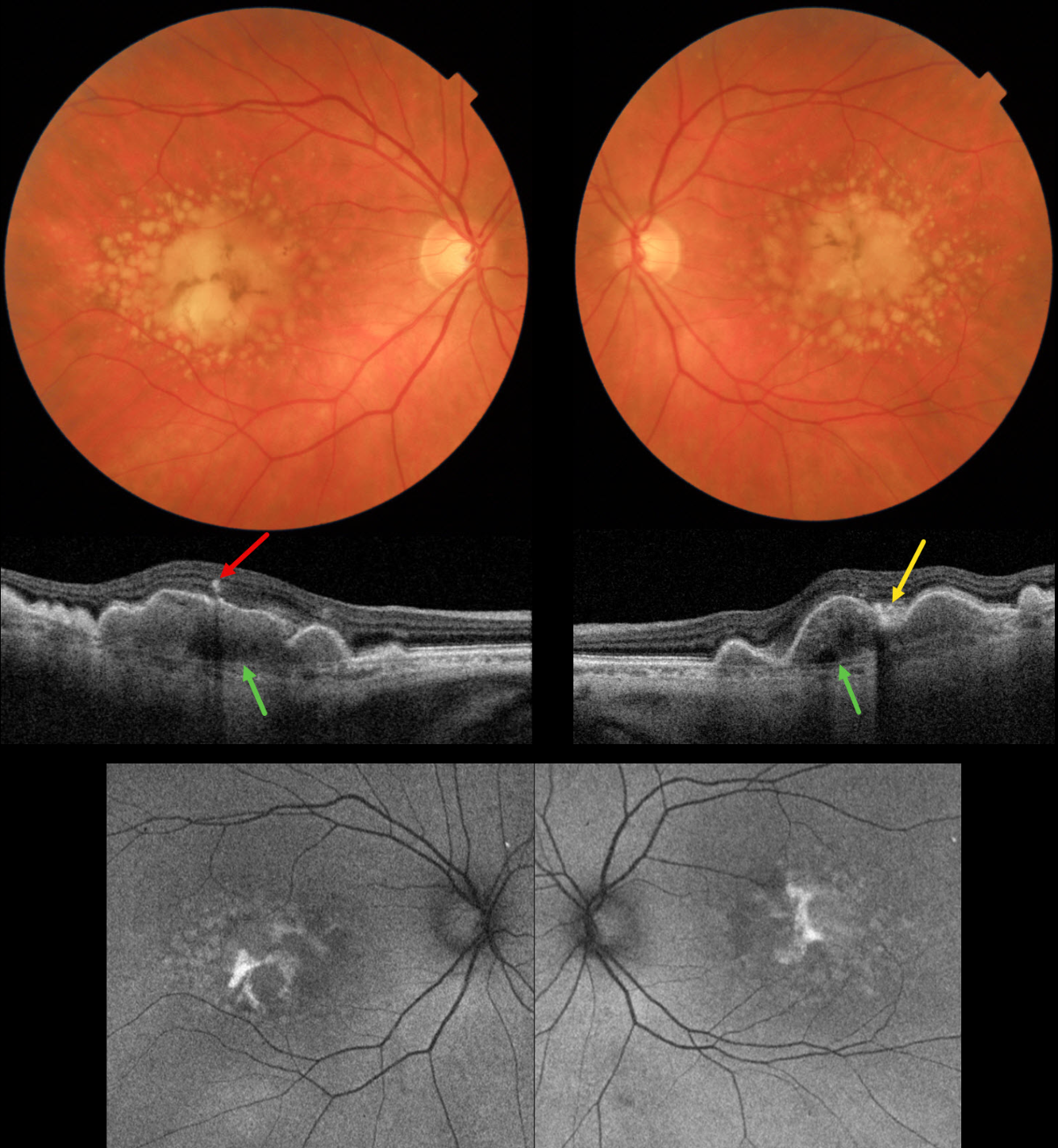
This 72YO female presented for her 6-month AMD examination. She was visually asymptomatic, and vision was 20/40 OU.
Triton color imaging shows bilateral chronic foveal RPE detachments (PEDs) with surrounding soft drusen. A radiating pigment figure overlies each PED. Swept-source OCT shows hyperreflective material beneath each PED, with some overlying hyperreflective foci and hyperreflective material likely representing lipofuscin. Bruch membrane is visible beneath the PED and surrounding soft drusen. Fundus autofluorescence (FAF) shows variable hyper-FAF from the pigment figures and lipofuscin.
Learning Points:
RPE detachments are most commonly associated with AMD and the central serous-pachychoroid spectrum. In cases of AMD, lesions can be avascular (as in our patient) or vascularized as part of a type 1 neovascular complex (below the RPE). AMD-related PEDs can form primarily or as a confluence of soft drusen. When they collapse, they may lead to secondary macular atrophy (Yu et al, Ophthalmology 2019;126:261-273).
The radiating pigment figure, described originally by Dr. Gass, is pathognomonic for a chronic PED and persists even after a PED flattens. Avascular PEDs are typically not treated. In contrast, anti-VEGF therapy is both safe and effective for vascularized PEDs, despite the risk of a RPE tear (Khanani et al, Retina 2018;38;2103-2117).



