

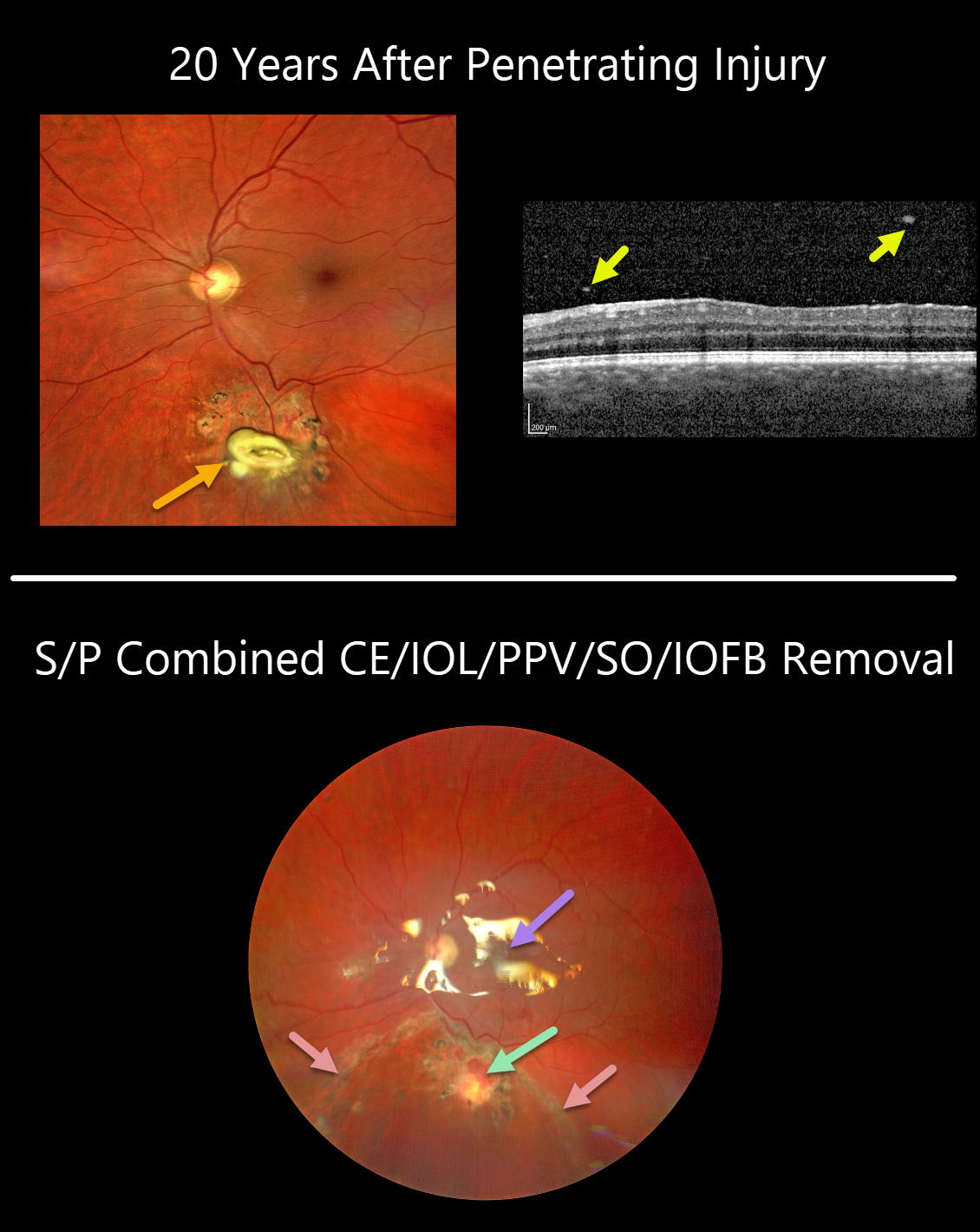
This 36YO male presented with recent blurred vision of 20/32 in his left eye.
Clarus imaging shows a whitish elevated lesion with surrounding chorioretinal scarring just below the inferotemporal arcade. OCT scanning shows overlying vitreous cells.
He gave a history of a retained retinal plastic foreign body 20 years earlier, following a penetrating injury from an exploding detonator. He had no prior visual complaints, and given the presumed inert nature of the intraocular foreign body (IOFB), surgery was not recommended at that time.
Combined phacoemulsification and vitrectomy with foreign body removal was performed due to increasingly symptomatic vitritis. The retina around the IOFB was necrotic and excised, and the area was surrounded with laser retinopexy. The retina was completely attached under silicone oil one week later.
After removal, the central portion of the IOFB was found to be metallic but coated in plastic. Presumably, as the plastic degraded over time, the metal’s toxicity caused secondary inflammation and visual symptoms.
Learning Point:
In retrospect, a CT scan should have been performed following the initial injury to ensure that the IOFB was fully non-metallic.



