

This 34YO male presented with pain and vision loss immediately following a metal-on-metal injury at work. Vision was light perception. Slit lamp examination revealed a corneal laceration and traumatic cataract. B-scan ultrasonography and CT scanning revealed an intraocular foreign body (IOFB). The patient was immediately taken to surgery for corneal wound repair, lensectomy, and vitrectomy.
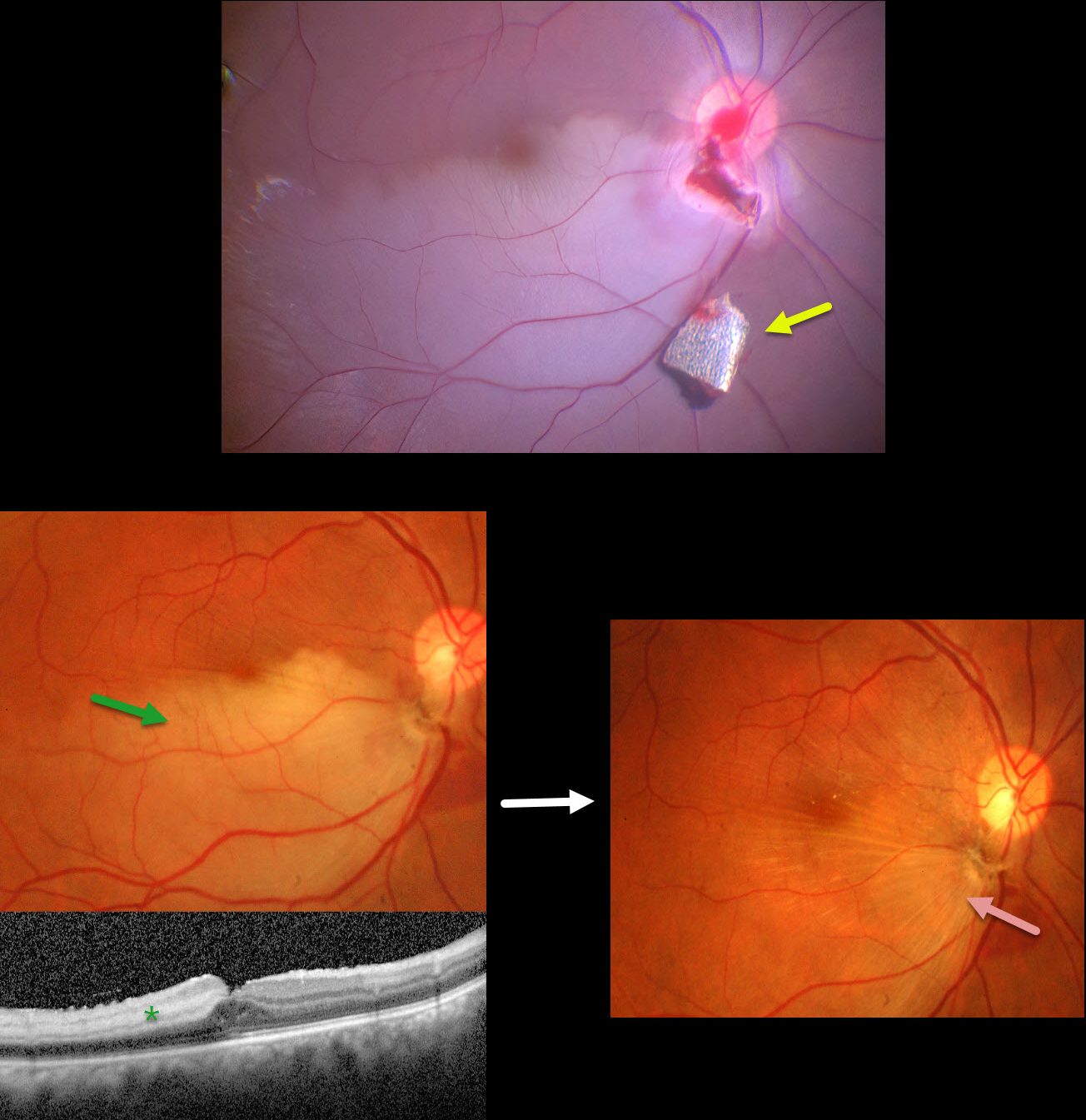
Intraoperative photography shows a metallic intraocular foreign body (IOFB) resting just inferior to the optic nerve. The impact site is just inferior to the optic nerve, with an inferotemporal branch retinal artery occlusion (BRAO) distally. The IOFB was removed with a rare-earth magnet.
Two weeks following surgery, vision was 20/120 with an aphakic correction. The inferior retina remains opaque, and the OCT shows inner retinal hyperreflectivity from the BRAO. A secondary intraocular lens implantation was performed 3 months postoperatively. Eleven months following the initial surgery, the BRAO is fully resolved, and secondary retinal striae extend outwards from the impact site. Vision remarkably improved to 20/30.
This case was published in the Indian Journal of Ophthalmology (Nagpal M et al, Ind J Ophthalmol 2018;66;146-148).



