

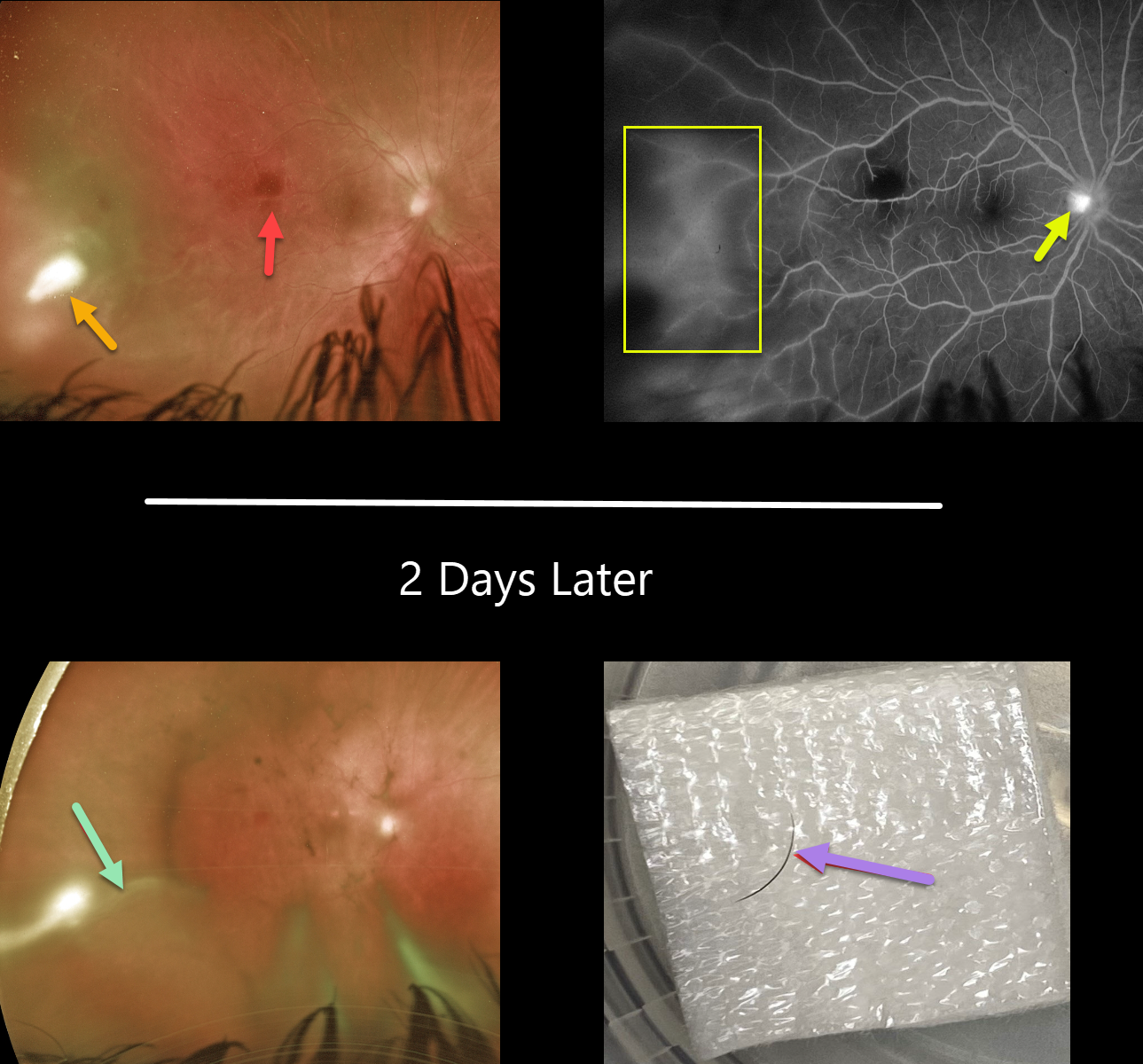
This 18YO female presented with a 10-day history of pain, redness, and blurred vision in her right eye. Her referring doctor put her on topical prednisolone acetate 1% and ciprofloxacin several days earlier.
Vision was 20/60 OD and 20/20 in her normal left eye. There was significant temporal conjunctival injection overlying a deeper scleral nodule with moderate chemosis. There were fine KP on the corneal endothelium, severe cell and moderate flare, and a 0.8mm hypopyon with scattered fibrin clumps (not shown).
Fundus examination shows an outer retinal hemorrhage in the distal macula, along with a patch of white retinal/vitreous inflammation in the temporal periphery. Fluorescein angiography shows diffuse leakage temporally, along with some optic nerve leakage.
She was placed empirically on oral fluconazole. Laboratory workup for various infectious (toxoplasmosis, bartonella, syphilis, tuberculosis, toxoplasma) and inflammatory (Behcet’s, HLA-B27, ANA, ESR) causes was ordered.
One week later, vision was stable, and the scleral and anterior chamber inflammation were only minimally better. Given the lack of significant improvement, oral Bactrim was started.
She returned 2 days later with increasing vitreous inflammation and elongation of the temporal white vitreous opacity. Laboratory results were completely negative. She was taken to surgery the following day to both clear the visual axis and to obtain vitreous for cultures and sensitivities.
During core vitrectomy, the vitreous was so thick that the cutter had to be pulsed on and off due to the tip being occluded. As the elongated vitreous opacity was excised, an embedded underlying eyelash cilia was discovered. The lash was ‘plucked’ with intraocular forceps and removed from the eye, and the site was prophylactically surrounded with laser.
One week later, vision improved to 20/30. The external and intraocular inflammation were dramatically improved. However, a new superotemporal dialysis with peripheral retinal detachment was found, which was repaired with vitrectomy.
Learning Points:
This is an unbelievable case, and it is difficult to fathom how the eyelash entered the vitreous cavity. There was no history of trauma or ocular surgery.



