

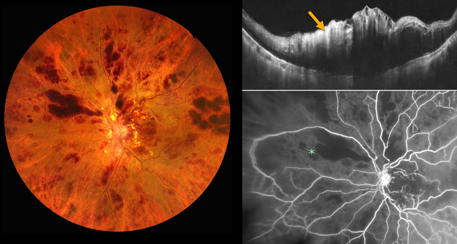
This 78YO male presented with 1 week of sudden, painless decreased vision in his left eye. Vision was 20/20 in his normal OD and light perception OS. Despite iris neovascularization, intraocular pressure was normal.
Color photography shows a hemorrhagic central retinal vein occlusion (CRVO) with variably confluent inner retinal hemorrhages and scattered nerve fiber layer infarcts. OCT scanning shows marked macular thickening with a hyperreflective inner retina. Fluorescein angiography shows profound diffuse retinal capillary loss. An intravitreal anti-VEGF injection was given. One week later, the rubeosis completely regressed, and full panretinal photocoagulation was performed.
Learning Points:
Ischemic CRVO, which accounts for 20% of eyes, is characterized by severe visual loss (typically worse than 20/200), a relative afferent pupillary defect, extensive retinal hemorrhages, cotton-wool spots, marked venous dilation and tortuosity, optic disc edema, and large areas of angiographic nonperfusion of the retinal capillaries.
Untreated iris neovascularization usually leads to neovascular glaucoma (NVG). Treatment involves an immediate anti-VEGF injection to allow for more rapid regression of neovascularization, followed by full panretinal photocoagulation. NVG is much less common in the modern anti-VEGF era, since the majority of these eyes also have macular edema that requires ongoing anti-VEGF therapy.



