

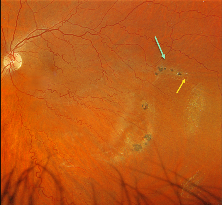
This 42YO male was referred for asymptomatic findings on retinal examination in his left eye. Vision was 20/20 in his normal OD and 20/20 OS.
Optos color RGB imaging shows patches of variably pigmented lattice degeneration in the inferotemporal midperiphery. At least one atrophic retinal hole is noted in the superior-most lesion.
The retinal veins are also diffusely tortuous except superotemporally, likely indicating a prior spontaneously resolved retinal vein occlusion.
Observation was recommended.



