

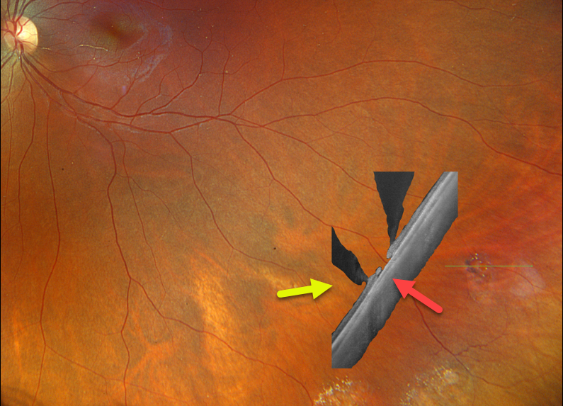
This healthy 27YO female has a known history of lattice degeneration with atrophic holes. She is completely asymptomatic, and vision is 20/20 OU.
Optos color RGB imaging of her left eye shows patches of inferotemporal lattice degeneration. Optos California Silverstone swept-source OCT through a lesion containing an atrophic retinal hole reveals classic findings, including retinal thinning within the lattice, liquified vitreous overlying the lattice lesion, and adherent vitreous along the edges of the lattice.
Learning Points:
Lattice lesions have a pocket of vitreous liquefaction overlying the thinned retina with a firm vitreo-retinal adhesion along their borders. White, fibrosed vessels often crisscross the lesions, which leads to the name “lattice”. This firm vitreoretinal adhesion, along with the often present atrophic holes, is what can predispose these eyes to retinal detachment.



