

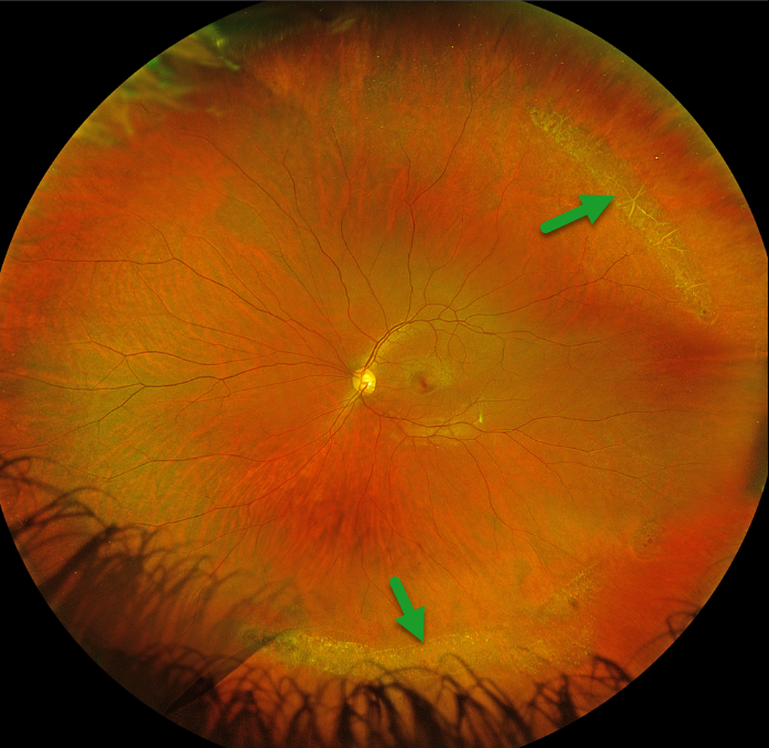
This 35YO female was referred for asymptomatic lattice degeneration. Vision was 20/20 OU.
Optos color RGB imaging of her left eye shows prominent temporal and inferior lattice lesions, some of which are associated with atrophic retinal holes. Similar findings were noted in her right eye (not shown).
Learning Points:
Lattice lesions have a pocket of vitreous liquefaction overlying the thinned retina with a firm vitreo-retinal adhesion along their borders. White, fibrosed vessels often crisscross the lesions, which leads to the name “lattice”. These are particularly well seen in our patient’s superotemporal lesion. This firm vitreoretinal adhesion, along with the often present atrophic holes, is what can predispose these eyes to retinal detachment.
Retinal detachment occurs in 1 in 10,000 people per year. Lattice is found in 10% of the general population, especially in high myopes. Since about one-third of patients with retinal detachment have lattice as the cause, the question always arises: Should prophylactic retinopexy be performed?
The short answer is that asymptomatic lattices generally should not be treated. To put things into perspective, imagine a sports stadium of 30,000 people. Three people will develop a retinal detachment in the coming year. Only one of the 3,000 people in the stadium with a lattice would develop one of those 3 detachments. Assuming prophylactic treatment would be 100% effective and complication-free, 2,999 eyes would be unnecessarily treated to prevent one detachment.



