

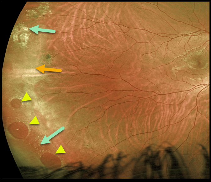
This 24YO male presented with asymptomatic but somewhat confusing clinical findings beautifully captured by this Optos image.
Three large atrophic retinal holes within an area of lattice are found in the inferotemporal periphery, along with a few areas of lattice and scarring superotemporally. There is also associated white without pressure, giving the appearance of a retinal detachment (remember the normally transparent retina turns whitish when it detaches). However, the retina was completely attached, and we are treating him with observation.
Learning Points:
Retinal detachment occurs in 1 in 10,000 people per year. Lattice is found in 10% of the general population, especially in high myopes. Since about one-third of patients with retinal detachment have lattice as the cause, the question always arises, especially in patients like this: Should prophylactic retinopexy be performed?
The short answer is that asymptomatic lattice generally should not be treated. To put things into perspective, consider a pre-COVID, non-social-distancing football stadium with 30,000 people. Three people will develop retinal detachment in the coming year. Only one of the 3,000 people in the stadium with lattice would develop one of those 3 detachments.
Excellent references on this controversial topic include: Byer Ophthalmology 1989;96;1396-1402, Folk et al Ophthalmology 1989;96;72-79, and Folk et al Retina 1990;10:165-169.



