

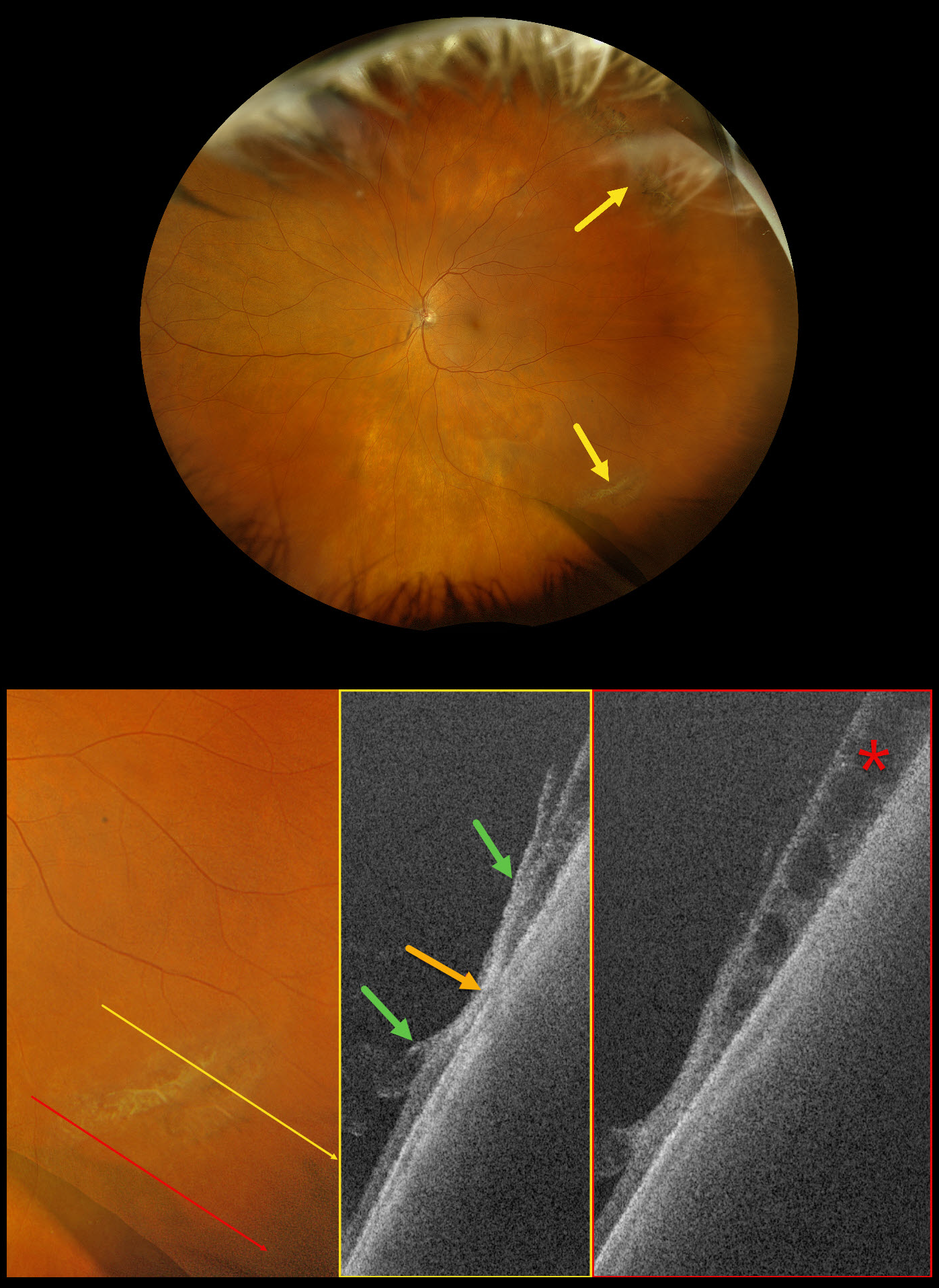
This 73-year-old female was referred for a symptomatic posterior vitreous detachment (PVD) with a retinal break in her left eye. Vision was 20/20.
Optos Silverstone RGB imaging shows supero- and inferotemporal lattice degeneration with some atrophic retinal holes. Swept-source OCT through the lattice shows a thinned, disorganized retina with areas of vitreoretinal adhesion along its margins. A more inferior line scan shows degenerative retinoschisis, which could not be visualized clinically or on the Optos image.
Our patient showed no retinal breaks from her symptomatic PVD, and observation was recommended.
Learning Points:
Ultrawidefield imaging has revolutionized our specialty, making many of us realize how limited our clinical examinations are compared with our imaging devices. In our patient’s case, the RGB image captured the fundus nearly from ora to ora. Swept-source OCT provided a near histologic view of the lattice pathology, including atrophic retina centrally, with a firm area of vitreoretinal adhesion along its margins. And finally, it revealed subclinical degenerative retinoschisis.



