

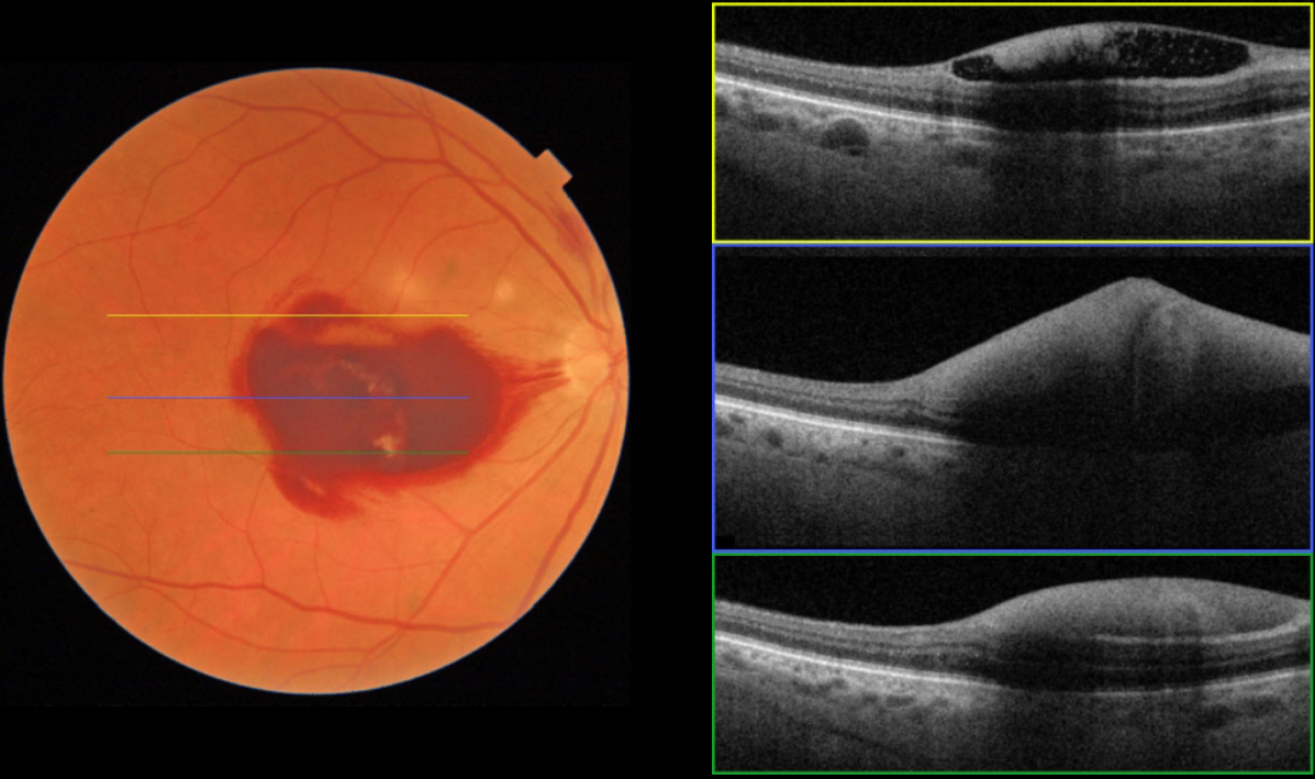
This 63YO male, who is undergoing treatment for leukemia, presented with one day of acute vision loss in his right eye. He was recently discharged from the hospital for chemotherapy-induced pancytopenia. His CBC showed: WBC 0.5, HgB 5.6, and Platelets 2. Vision was counting fingers OD.
Fundus imaging shows sub-internal membrane (ILM) hemorrhage. The OCT scans confirm hemorrhage in the sub-ILM space. We expect our patient’s macular hemorrhage to resolve as his blood counts normalize
Learning Points:
Ocular leukemic manifestations are primarily caused by accompanying hematologic abnormalities, including hyperviscosity, anemia, and thrombocytopenia (Soman et al, Ophthalmology Retina 2018;2:17-23).
Posterior segment findings include retinal hemorrhages (sub-ILM, nerve fiber layer, or deeper, white-centered), cotton-wool spots, dilated retinal veins, and serous retinal detachment. Leukemic infiltration of the optic nerve, retina, and choroid can also occur.
The sub-ILM hemorrhages are usually self-limiting and resolve within several months of treating the underlying disease (Ophthalmology Retina 2018;2:494-501).



