

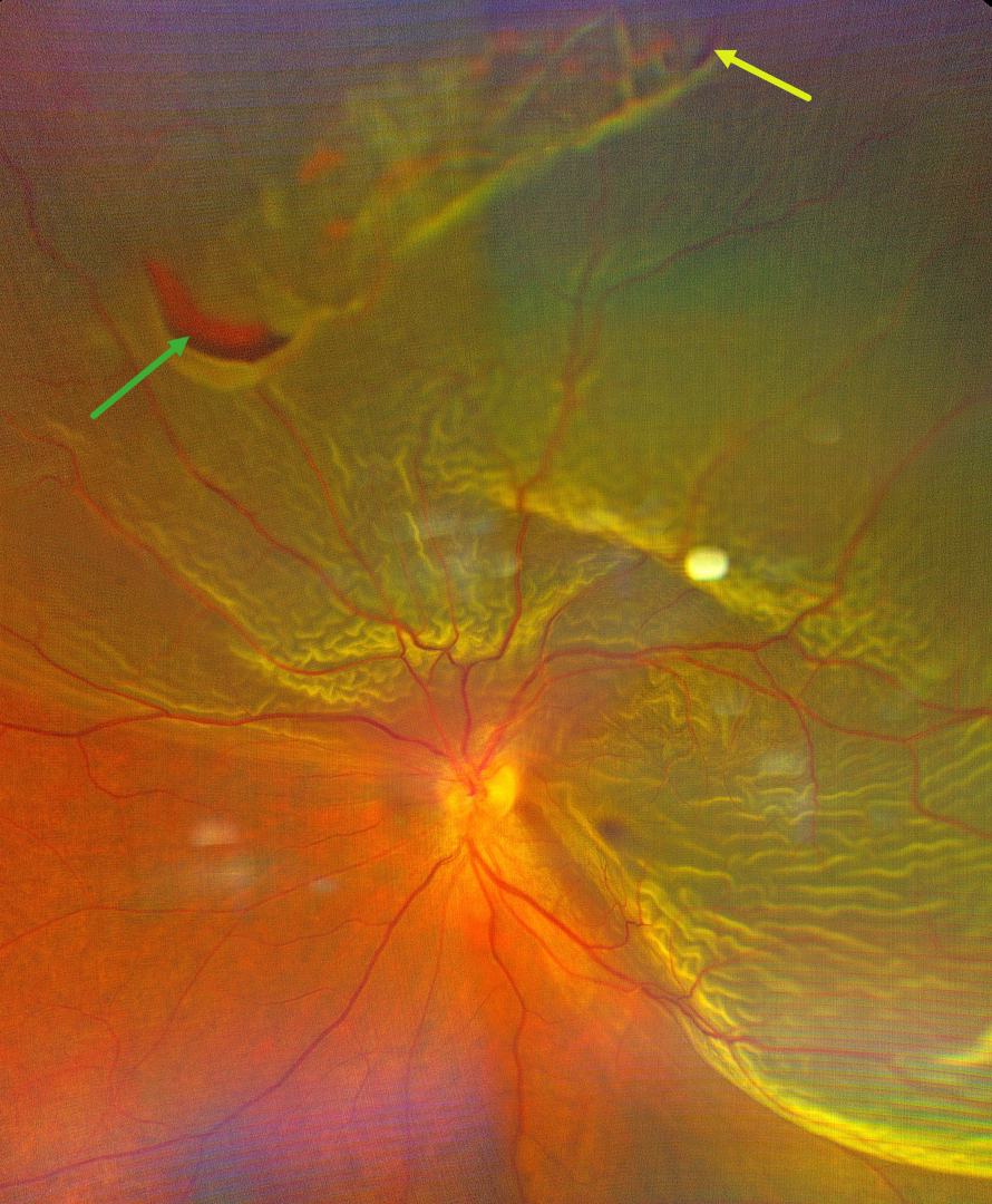
This 44YO male presented with sudden, painless, and progressive central and inferior peripheral vision loss. Color photography shows a superior and temporal macula-off rhegmatogenous retinal detachment (RRD). There is a superior patch of lattice degeneration with retinal tears at either edge of the lattice at 10:30 and 12:30.
Learning Points:
RRD occurs when liquid vitreous accesses the subretinal space through at least one full-thickness break in the neurosensory retina. A key principle for surgical success is identifying and closing all breaks. In 1971, Harvey Lincoff, in his classic paper ‘Finding the Retinal Hole,’ defined 4 findings based on RRD morphology that guide clinicians in determining where the causative breaks should be found (Lincoff and Gieser, Arch Ophthalmology 1971;85:565-569). These have since become known as the ‘Lincoff Rules.’ Simply put, gravity helps explain a detachment’s configuration and the location of the causative breaks.
Lincoff Rule 2 specifically addresses superior detachments crossing the 12 o’clock midline. It states that the primary break lies within a triangular zone with an apex at 12 o’clock, spanning 1.5 clock hours on either side (10:30-1:30 positions). This rule applies to 93% of such cases, as gravity causes subretinal fluid to spread symmetrically downward from the break.
Our patient’s detachment extends more inferiorly on the temporal edge. The two identified breaks in our image probably do not fully explain our RRD’s configuration, since the inferior edge of the detachment should fall on the nasal, not the temporal edge. At surgery, we would therefore search extra hard for at least one additional break along the temporal side.



