

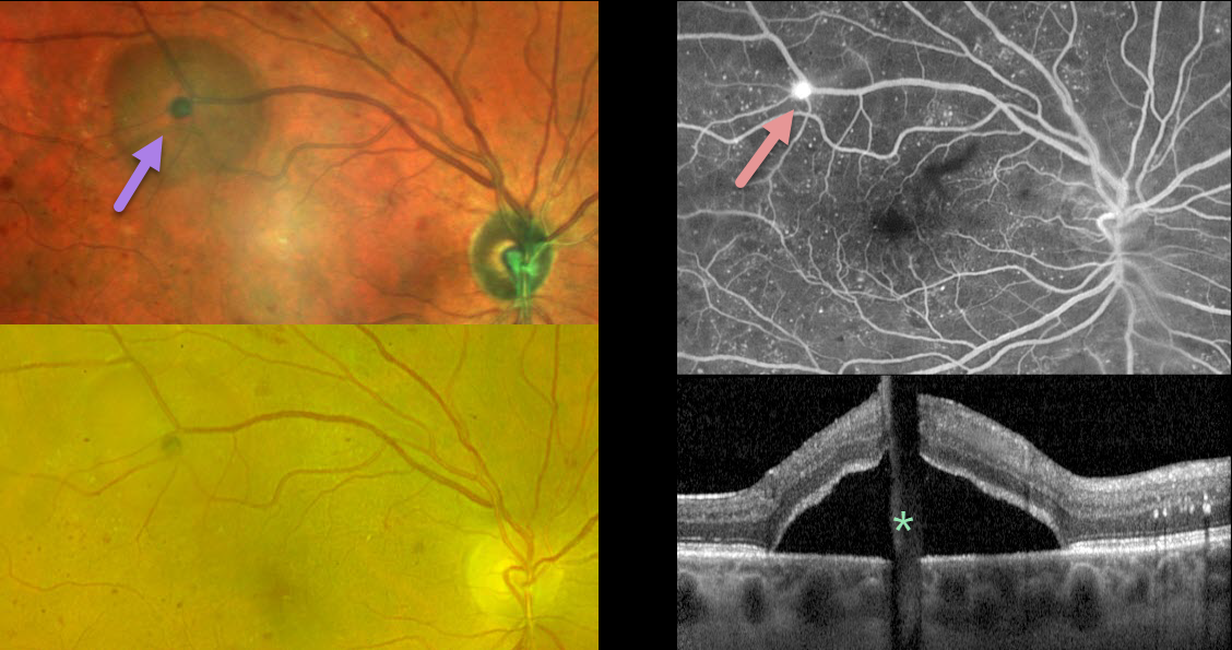
This 55YO female with type 2 diabetes presented for a routine follow-up without visual complaint. Vision was 20/30 bilaterally.
MultiColor imaging of her right eye shows a localized traction retinal detachment (TRD) in the distal superotemporal macula. The TRD is much less obvious with Optos color RG imaging.
The source of the traction is a small area of retinal neovascularization that leaks on fluorescein angiography. This localized traction is also confirmed on OCT. Although vitrectomy was not recommended given the localized nature of the TRD, panretinal photocoagulation was performed.
Learning Points:
Although pretty color images that match what we see on biomicroscopy are most intuitive, newer imaging modalities that ‘distort’ the normal fundus colors often contain very clinically relevant information. Standard Optos color RG images are generated from a red (635nm) and green (532nm) laser. Although these images have a greenish tint, they essentially contain two images in one: the RPE/neurosensory retina is best captured with the green channel, and the choroid is best seen with the red channel. A newer Optos true-color RGB unit has recently been introduced, which maintains the ability to view the separate RG channels while also generating a true-to-life color image.
Heidelberg Engineering’s Multicolor imaging module similarly utilizes a blue (488nm), green (515nm), and infrared (820nm) confocal laser to generate a fundus image. The blue laser best images the vitreoretinal interface and inner retina; the green laser images the mid retina; and the infrared laser images the outer retina, RPE, and choroid (Roy et al., Surv Ophthalmology 2024;69:378-402).
Combining these 3 wavelengths into a single file produces a pseudocolor image that, like the Optos, provides the clinician with more information than a standard color photo. As seen in our patient, Multicolor imaging best illustrates both the presence and extent of TRD (Gadde et al, Indian J Ophthalmol 2022;70:465-470).



