

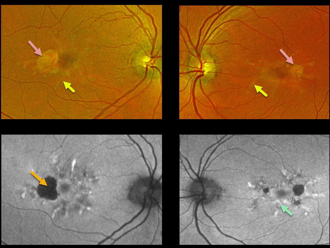
This 62YO female presented with 20/20 vision OU and no visual symptoms. Optos imaging shows bilateral radiating subretinal yellowish macular lesions with temporal atrophy.
Fundus autofluorescence (FAF) shows variable hyper-FAF from these lesions, along with hypo-FAF within the areas of atrophy.
Learning Points:
Pattern macular dystrophies are associated with numerous mutations, including ABCA4, PRPH2, BEST1, PROM1, CERKL, CNGB3, and CRB1, among others (see Del Pozo-Valero et al, Invest Ophthalmol Vis Sci 2022;63(2):11).
These patients tend to maintain fairly good vision, although outer retinal and RPE atrophy can cause central vision loss, as can the rare development of macular neovascularization. Patients should therefore regularly monitor their vision with an Amsler grid.



