

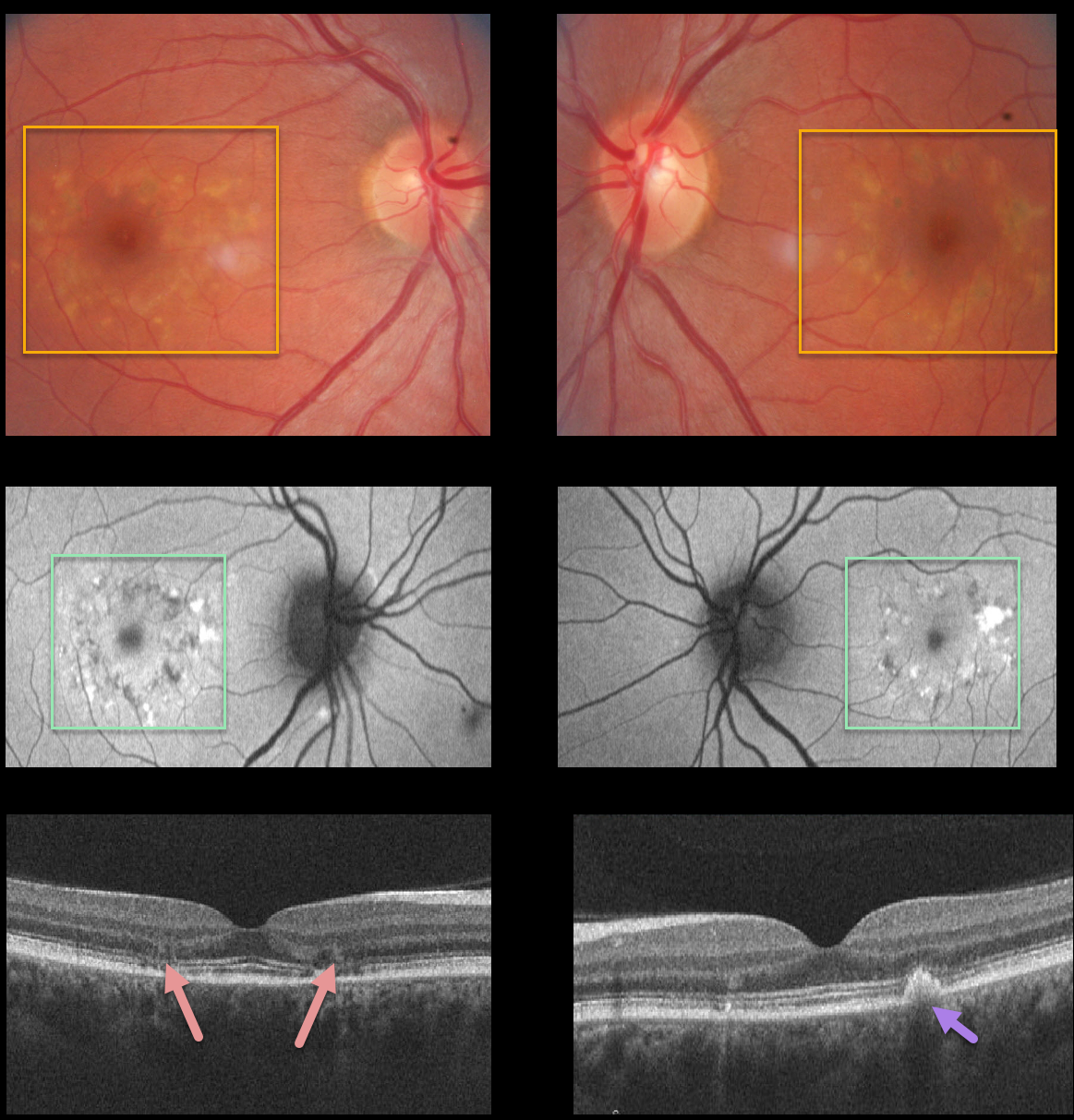
This 34YO female, a local optometrist’s sister, was referred for these asymptomatic bilateral macular changes consistent with a macular dystrophy.
Fundus images show symmetrical yellowish subretinal pigmented drusen-like changes. Fundus autofluorescence reveals that these lesions are variably hyper- and hypoautofluorescent.
OCT scans show some variable ellipsoid zone loss and outer retinal atrophy, as well as subretinal hyperreflective material. Her brother had similar findings. Unfortunately, we have no further clinical information.
Learning Points:
Pattern macular dystrophy is a group of disorders characterized by pigment deposition of the macula and caused by mutations in the RDS/peripherin gene. The following are considered types of pattern dystrophies: adult-onset foveomacular vitelliform dystrophy, butterfly-shaped pigment dystrophy, reticular dystrophy, multifocal pattern dystrophy simulating Stargardt disease, and fundus pulverulentus.
Pattern macular dystrophy has also been found in systemic disorders such as pseudoxanthoma elasticum, myotonic dystrophy, and McArdle Disease.



