

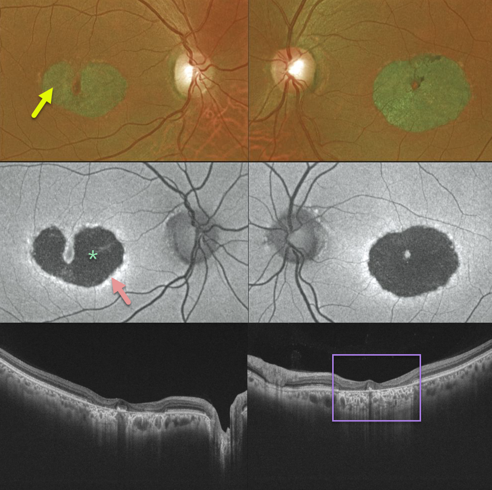
Our patient has a bilateral bull’s-eye maculopathy with central foveal sparing. Vision was 20/30 OD and 20/60 OS.
Fundus autofluorescence shows central hypo-FAF from loss of the central RPE. The thin surrounding hyper-FAF ring likely represents diseased, at-risk RPE cells. The OCT scans show pericentral outer retinal and RPE loss.
Learning Points:
Our patient no doubt has some type of inherited macular disorder. Some possibilities include central areolar chorioretinal dystrophy or a cone-rod dystrophy.
Central areolar chorioretinal dystrophy is a genetic condition associated with mutations in the RDS/peripherin gene, which encodes for a cell surface glycoprotein found in the rod and cone outer segments. The RPE and choriocapillaris atrophy over time, eventually forming a round, atrophic lesion in the macula. Vision can vary from 20/20 to 20/200.



