

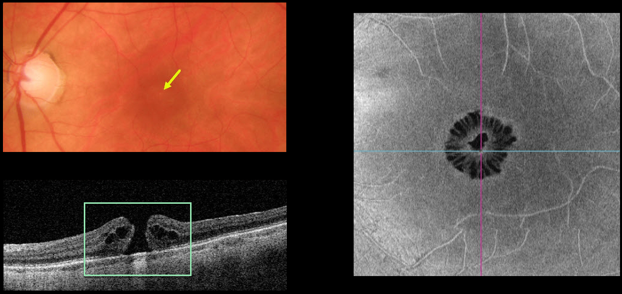
This 65YO female presented with 20/200 vision in her left eye from a symptomatic full-thickness macular hole. The hole is barely visible funduscopically.
Swept-source OCT shows the classic appearance of a hole from a foveal dehiscence with cystic thickening. En face imaging shows a unique and beautiful perspective of the hole.
Learning Points:
As described by Govetto et al (AJO 2020;212:43-56), idiopathic macular holes have a sunflower appearance showing features of both exudative and tractional cystoid spaces.
The macular hole is hyporeflective centrally with multiple hyporeflective cystoid spaces radiating from the center.



