

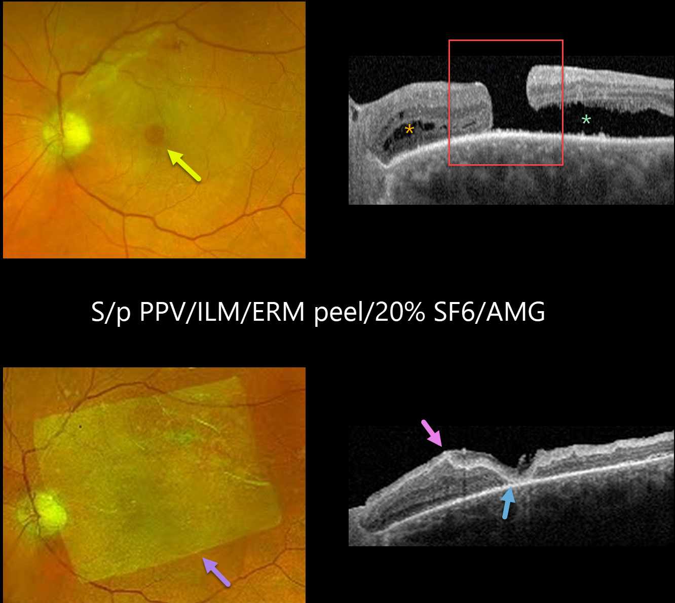
This 77YO male had a history of failed vitrectomy with internal limiting membrane (ILM) peeling and SF6 gas for a long-standing full-thickness macular hole, and presented for a second opinion. Vision was 20/125.
Optos color RG imaging shows a persistent macular hole with some underlying subretinal fluid. There is a residual epiretinal membrane (ERM) along the superior arcade.
OCT shows the full-thickness macular hole with outer retinal cysts nasally and subretinal fluid temporally. Some ellipsoid zone (EZ) and outer retinal changes are visible.
The patient was eager to try additional surgery and underwent a vitrectomy with ERM/ILM peel, endolaser, 20% SF6, and amniotic membrane graft (AMG) placement. At 9 weeks post-op, the patient’s vision had improved to 20/80.
Fundus imaging shows a sheet of AMG over the macular hole and several superior laser scars. OCT scanning shows the macula hole to be closed under the AMG with EZ and outer retinal changes, but resolved temporal subretinal fluid.
Learning Points:
Approximately 90% of macular holes close with a single surgery. However, larger holes over 400 microns and more chronic holes can be challenging to close. Several techniques have been described to treat these refractory holes, including a broader ILM peel, instruments such as a Flex Loop to loosen up the edges of the hole, amniotic membrane grafts, or various variations of inverted ILM flaps to drape the hole, and even autologous full-thickness retinal tissue transplantation.



