

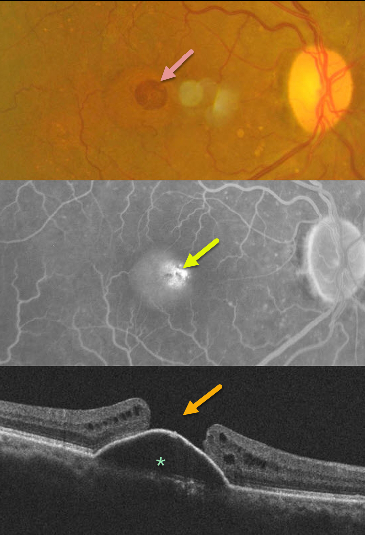
This 60YO female presented with 1 month of vision loss in her right eye. Vision was 20/100 OD and 20/40 OS.
Fundus photography of her right eye shows a large macular hole with some nasal pachydrusen. There was an old branch retinal vein occlusion without macular drusen in her left eye (image not shown).
Fluorescein angiography shows central pooling beneath a retinal pigment epithelial detachment (PED) with increased transmission through the macular hole. OCT scanning shows a PED with an overlying large macular hole.
This case presents a unique and interesting challenge in that macular hole surgery likely would not close the hole, given the underlying PED. She received 3 monthly Lucentis injections in her right eye, with no change in the PED.
Our thought would be to try a different anti-VEGF agent, such as Eylea, or consider adding photodynamic therapy for a possible pachychoroid-related avascular PED.
A similar case showed spontaneous closure of a macular hole following anti-VEGF therapy (see Chakraborty and Sheth, Indian J Ophthalmol Case Rep 2023;3:439-440).
If our patient’s macular hole remained open once the PED flattened, then macular hole surgery would be performed.



