

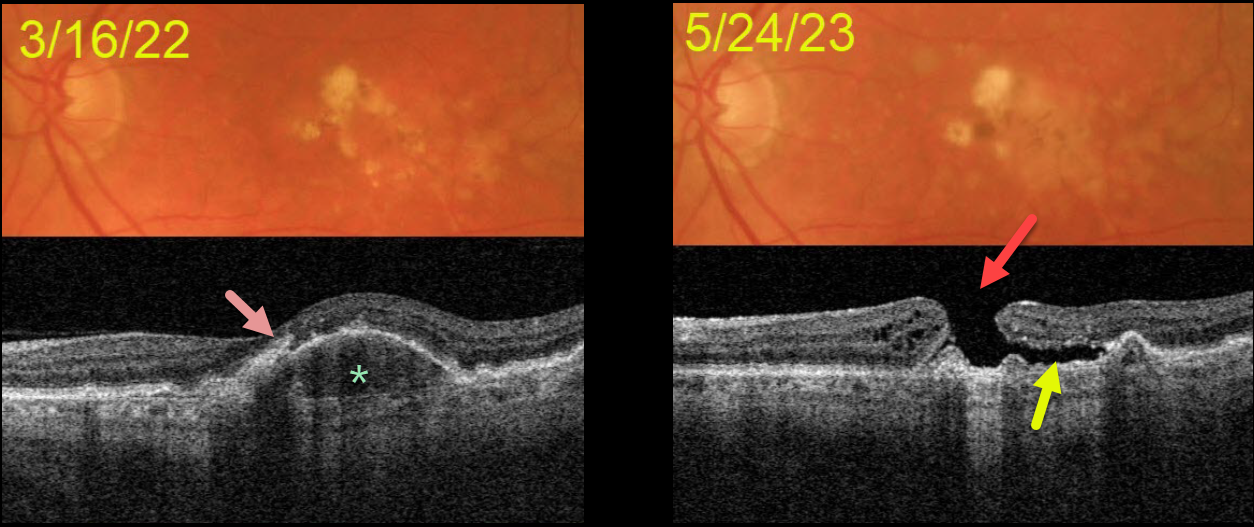
This 72YOM was examined with asymptomatic intermediate AMD bilaterally. Vision was 20/40 OD and 20/50 OS.
Color imaging of his left eye initially shows mostly large, soft drusen and pigment clumps. Swept source OCT shows a RPE detachment (PED) with overlying hyperreflective foci.
He returned over a year later without new symptoms, despite vision dropping to counting fingers. OCT shows a large macular hole overlying the collapsed PED. Shallow subretinal fluid lies below the temporal aspect of the hole.
The development of a macular hole following spontaneous collapse of a PED is quite unusual. Fortunately, vision in his right eye remains at 20/40 with intermediate dry AMD.
Since our patient was completely asymptomatic, he did not want macular hole surgery, and we will monitor him twice yearly.



