

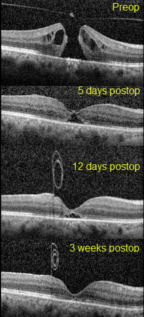
This 51yo presented with 20/1250 vision in her right eye from a primary full-thickness macular hole without traction measuring 407 microns. There was also a moderately severe nuclear sclerotic cataract.
Combined cataract surgery with 23-gauge pars plana vitrectomy, temporal half-moon inverted ILM flap, and fluid-air-SF6 exchange was performed, followed by prone positioning.
On the 5th postoperative day, the flap extended over the closed hole, but one week later, the ILM flap retracted and was rolled inward, suspended anterior to the macula.
Three weeks following vitrectomy, the hole was completely flattened with partial restoration of the outer retinal layers. The ILM flap continued to contract and scroll onto itself. Vision was 20/40.
Learning Points:
Introduced by Kelly and Wendel (Arch Ophthalmol 1991;109:654-659), macular hole surgery is one of the modern success stories in ophthalmology. ILM flaps appear to improve the surgical success for large (>400 microns) macular holes.
In this case, the ILM flap inadvertently scrolled onto itself and away from the macular hole by post-op day 12. Some authors have suggested that the ILM flap provides a scaffold for cells to grow over and help heal the hole.
However, most macular holes are actually foveal dehiscences without tissue loss, where the foveal retina splits and elevates like a drawbridge (Tornambe, Retina 2003;23:421-424). For successful surgery, there is thus no need or significant evidence for glial or neurosensory retina regrowth, just for the “drawbridge” to close.
As this case exemplifies, the ILM flap most likely serves the same purpose as the gas bubble, helping to sequester the macular hole from the overlying vitreous. This allows the RPE to actively pump (and the choroid to passively absorb) the subretinal fluid, allowing the foveal tissue to flatten into its normal position. Once the hole closes, as long as the vitreous and epimacular traction are relieved, there is likely no need for the ILM flap.



