

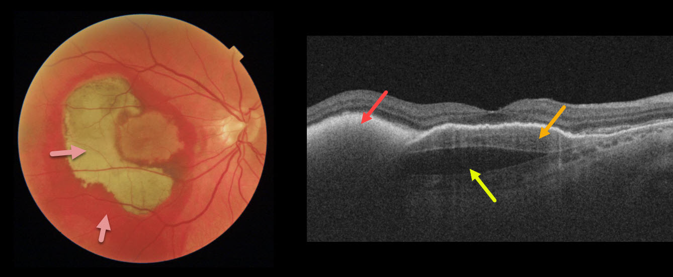
This patient’s vision remained 20/100 one month following vitrectomy with subretinal tissue plasminogen activator.
Surgery accomplished its goal of displacing most of the subfoveal blood, which is variably red and devitalized (yellow).
OCT shows the remaining subretinal hyperreflective blood, the causative sub-RPE type 1 macular neovascularization, and a subfoveal prechoroidal cleft.
Learning Points:
A prechoroidal cleft is a hyporeflective space between the RPE and Bruch’s membrane.
These are seen in up to about 20% of treated eyes with wet AMD, particularly with type 3 neovascularization and polypoidal choroidal vasculopathy.
They are usually associated with worse visual acuity due to potential complications, including RPE tear and subretinal hemorrhages.
For a more detailed discussion of prechoroidal clefts, see Kim et al Retina 2017;37;2047-2055.



