

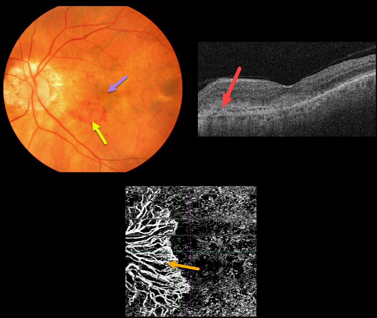
This 86YO male presented with symptomatic macular neovascularization (MNV).
Color imaging shows nasal patches of subretinal blood and more central subfoveal pigment. OCT scanning shows a hyperreflective MNV above the RPE (type 2 MNV). The peripapillary MNV is seen on OCT angiography.
Learning Points:
Macular neovascularization is also known by other terms, including choroidal neovascular membrane and subretinal neovascular membrane.
However, since these vessels are now known not to necessarily arise just from the choroid, the proper term is macular neovascularization (see Consensus on Neovascular Age-Related Macular Degeneration Nomenclature Study Group Ophthalmology 2020;127;616-636).
MNV is classified according to the location of the new vessels in relation to the RPE and neurosensory retina: type 1 (below the RPE), type 2 (between the RPE and neurosensory retina, and type 3 (within the neurosensory retina).



