

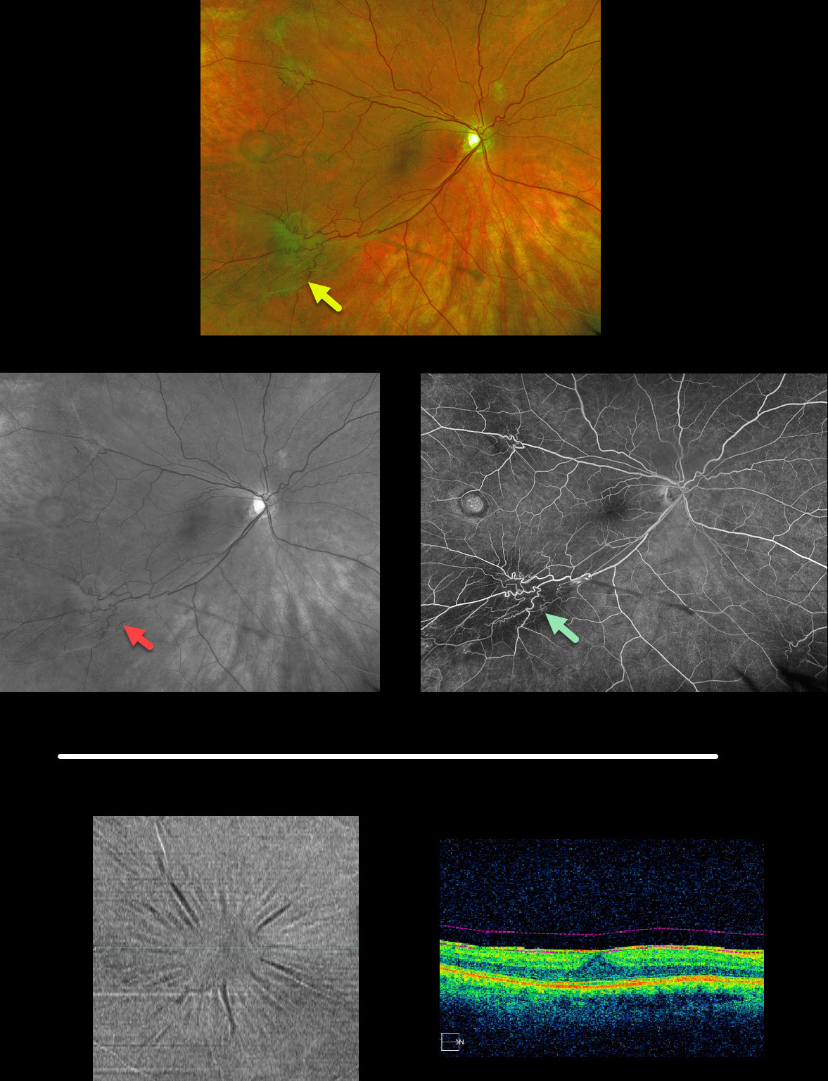
Multifocal images of an extramacular epiretinal membranes with an unrelated temporal retinal pigmented epithelial detachment are displayed here (Optos, red-free, and fluoroscein angiography).
BONUS: The second set of images is a different patient with a macular pucker that is well-visualized using OCT en face analysis, which provides transverse views of the retinal and choroidal layers using user-specified depths and thickness. This is an underutilized yet fantastic tool that gives another perspective for many chorioretinal disorders.



