

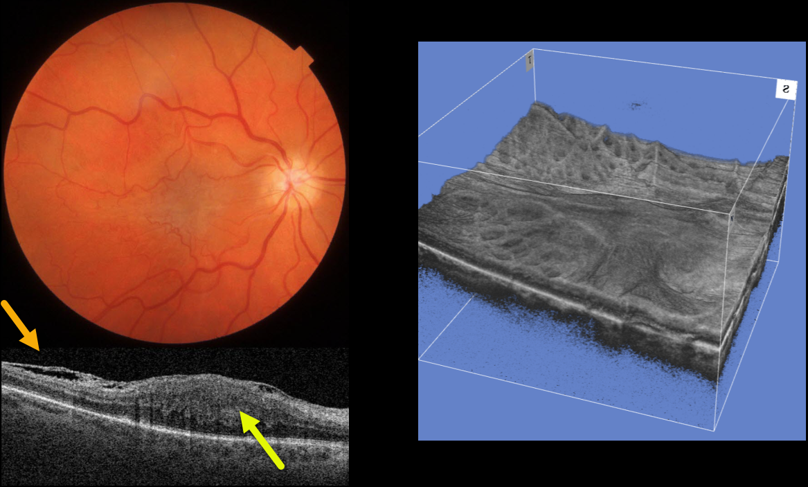
This 64YO female was referred for an asymptomatic macular pucker in her right eye. Vision was 20/200 OD and 20/20 OS.
Color imaging shows a severe central macular pucker. Triton swept-source OCT shows a variably adherent epiretinal membrane (ERM) with inner retinal hyperreflectivity and thickening, and complete loss of the normal inner retinal structures.
A 3D OCT reconstruction offers another view of the inner macular appearance, with unusual radiating dimples more peripherally.
Learning Points:
In 2017, Govetto et al described an OCT finding of ectopic inner foveal layers (EIFL) associated with epiretinal membranes (AJO 2017;175:99-113). The findings range from mild underlying retinal changes (stage 1, thin ERM with foveal depression), widening of the ONL and loss of foveal depression (stage 2), and ERM continuous with EIFL crossing the entire foveal area (stage 3). All retinal layers are clearly visualized in stages 1 through 3.
In stage 4, the ERM is thick, and all retinal layers are disrupted. Vision is worse with increasing stage severity. It is also associated with worse postoperative visual recovery (Yang et al, Retina 2022;42:1472-1478).
Combined hamartomas are classically described as benign congenital lesions composed of glial cells, vascular tissue, and pigmented retinal pigment epithelial cells. However, more recent data indicate that these lesions primarily arise from the inner retinal layers (see Chawla et al AJO 2017;181:88-96).
Combined hamartomas and severe macular puckers can look very similar, even to some of the most seasoned retina specialists. In our opinion, many cases of combined hamartomas in the literature most likely represent severe ERMs with EIFL.
Our patient has stage 4 EIFL. We recommended observation since she is binocularly asymptomatic.



