

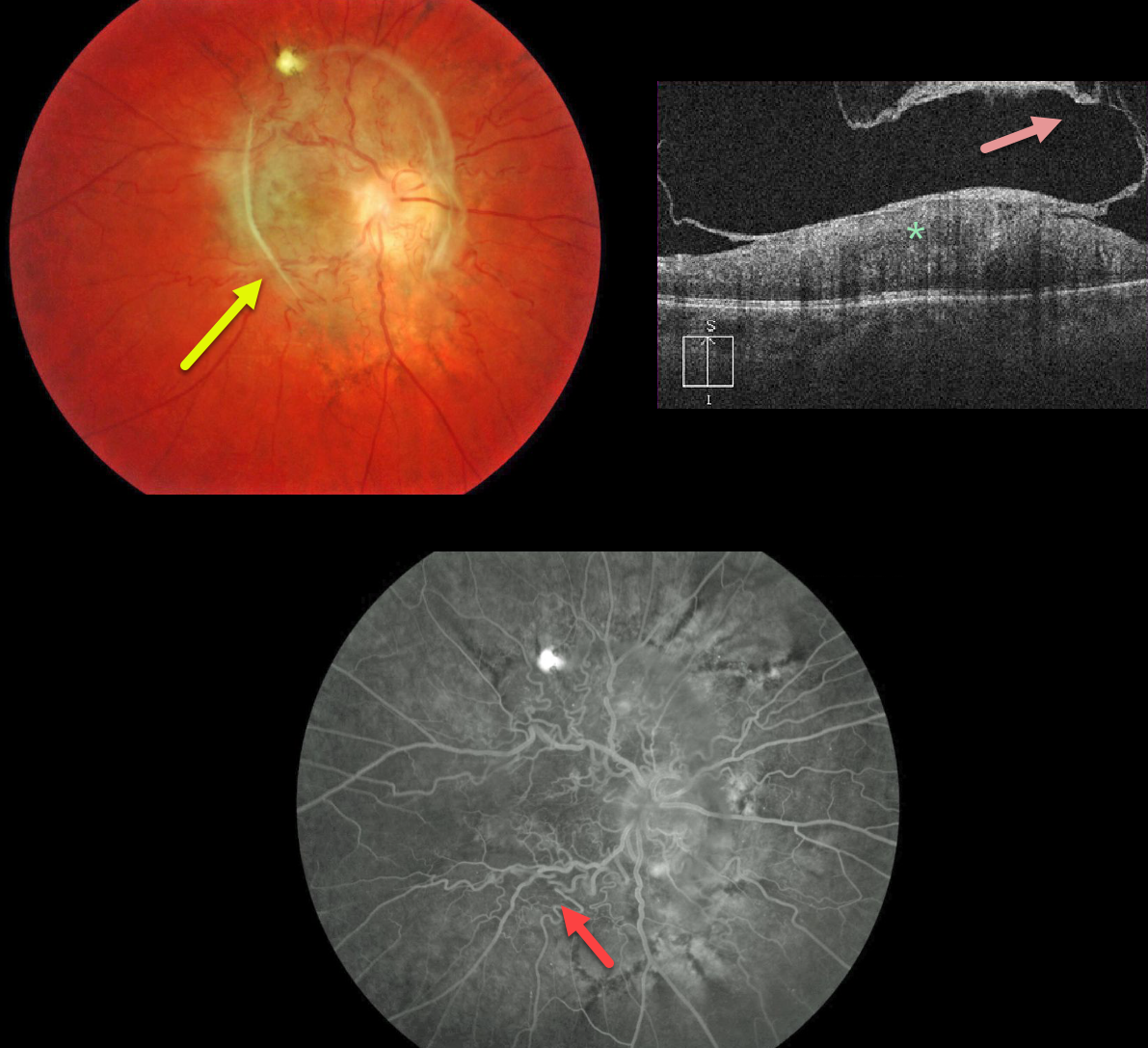
The fundus photo shows an unusual peripapillary pigmented epiretinal membrane distorting the underlying retina.
The OCT shows a partially detached epiretinal membrane and marked hyperreflectivity of the inner retina, with loss of normal retinal layer detail.
Fluorescein angiography shows a distorted retinal vasculature with late leakage.
Occasionally, vitrectomy with membrane peeling can be offered, but we are electing to observe this eye since vision has been poor since birth.
Learning Points:
A combined hamartoma of the RPE is a benign congenital lesion composed of glial cells, vascular tissue, and pigmented epithelial cells.
Even though we’re calling this a combined hamartoma, we’re not positive that this may not just be an unusual peripapillary/macular epiretinal membrane.



