

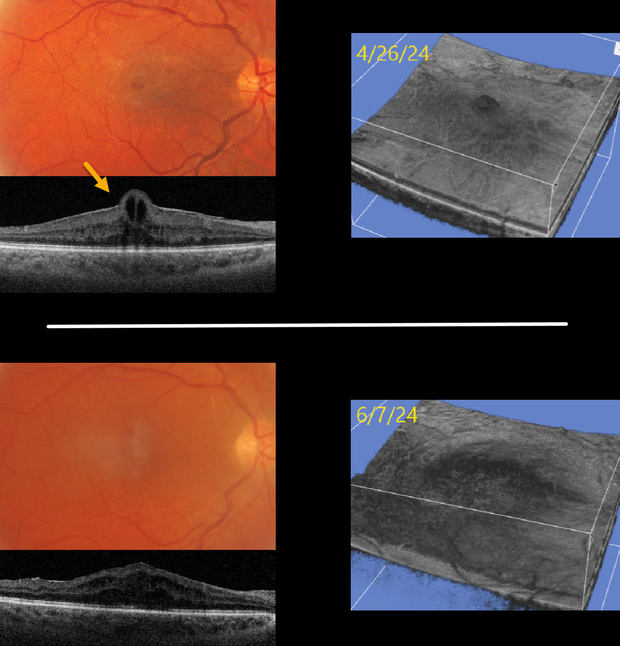
This 53YO male presented with counting-fingers vision in his right eye due to a symptomatic macular pucker.
Optos color RG imaging shows a macular pucker with a foveal cyst. Triton swept-source OCT shows an adherent pucker with central foveal herniation. Vitrectomy surgery was performed, and one month later, vision improved to 20/100. OCT scanning shows no pucker, moderate diffuse cystic retinal thickening, and resolution of the foveal herniation. The pre- and postoperative appearances are also well visualized with 3D OCT reconstruction.
Learning Points:
Foveal herniation is found in about 1% of eyes with idiopathic epiretinal membranes (Uzel et al, Eye 2023;37:1357-1360). Epiretinal membranes not uncommonly surround the foveal center, and central contraction can result in uninvolved inner foveal tissue being squeezed through the central membrane defect, resulting in this unique appearance. Vitrectomy surgery with membrane peeling usually results in si



