

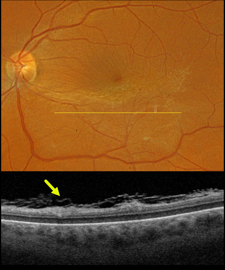
This 58YO female was referred for an asymptomatic macular pucker in her left eye. Vision was 20/25.
Optos color RGB imaging shows a moderate macular pucker centered in the inferior macula. Fine inner retinal striae are noted superiorly. Triton swept-source OCT shows a shredded appearance of multi-layered hyperreflective tissue overlying the retinal surface (the ‘spaghetti sign’).
Learning Points:
Fibrillary changes overlying an epiretinal membrane (ERM) were initially noted by Kim et al (AJO 2012;153:692-697), and termed schisis of the retinal nerve fiber layer by Hussnain et al (AJO 2019;207:304-3120). Despite rarely being described in the medical literature, it is present in over 50% of ERMs (Russell et al, Ophthalmology Retina 2023;7:325-332). Felt to arise from tangential tractional forces from the overlying epimacular tissue, it can appear clinically as whitish retinal areas overlying a pucker. During intraoperative membrane peeling, this tissue can be mistaken for residual epimacular proliferation and should not be removed to avoid iatrogenic retinal damage.



