

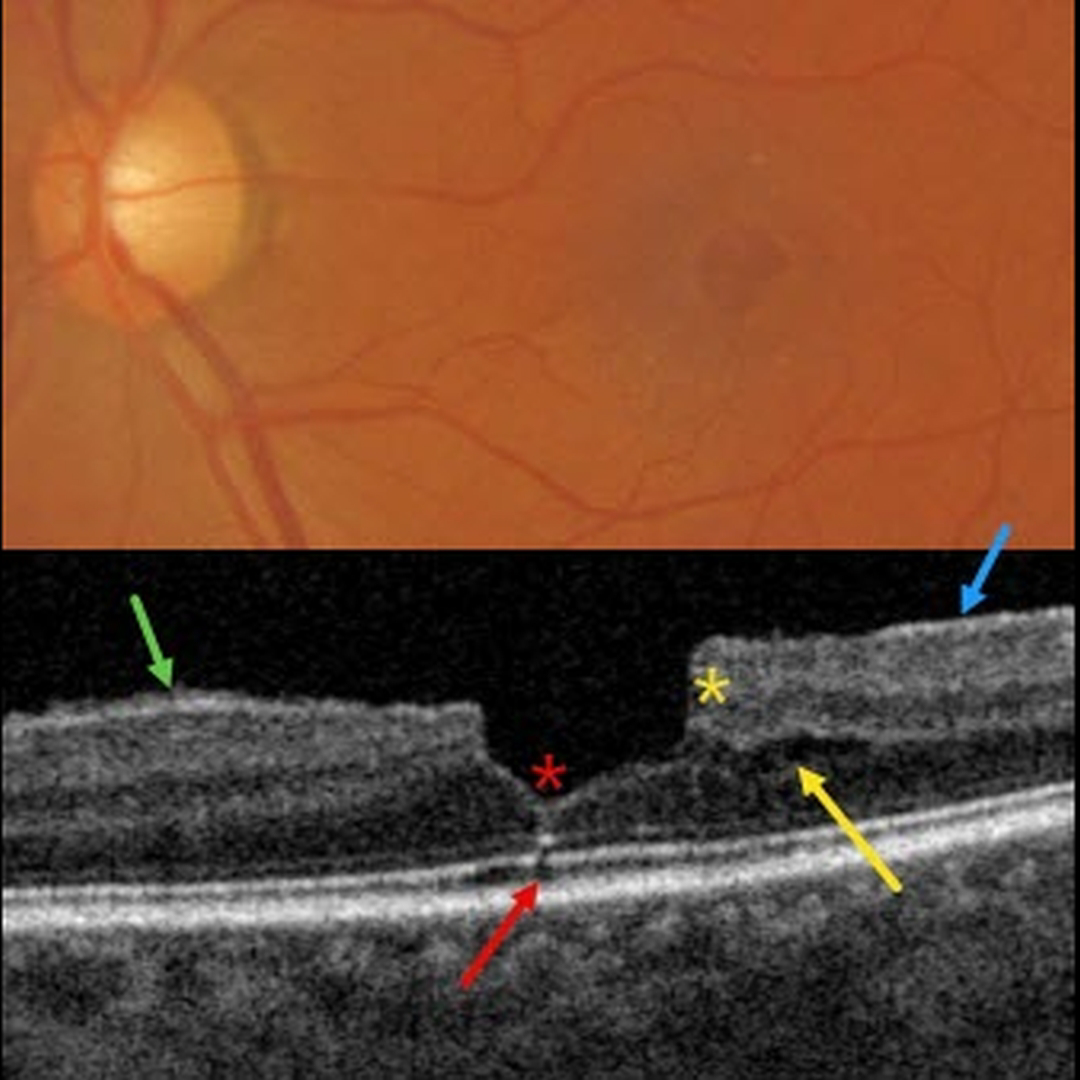
This patient presented with 20/30 vision due to a mildly symptomatic clinical macular pucker with a pseudohole; observation was recommended. Multimodal imaging reveals extensive pucker-related findings.
Triton color imaging shows an irregular macular pseudohole with a faint surrounding epiretinal membrane (ERM). True full-thickness macular holes are virtually always perfectly round, and these pucker-related pseudoholes are often irregularly round, as in this eye.
Swept-source OCT shows an adherent ERM with some shaggy-appearing adherent vitreous cortex, favoring epimacular proliferation. The fovea has a vertical contour, often present with ERM, due to the encircling membrane contracting towards the foveal center, dragging the inner retina centrally.
There are features of both a tractional lamellar macular hole (LMH) with splitting of the outer plexiform layer, along with a degenerative LMH with descending loss of tissue into the outer nuclear layer (Govetto et al, AJO 2016;164:99-109).
Finally, a tiny area of shadowing vs an outer foveal microdefect (Cohen et al, Ophthalmology Retina 2021;5:553-561) is seen.



