

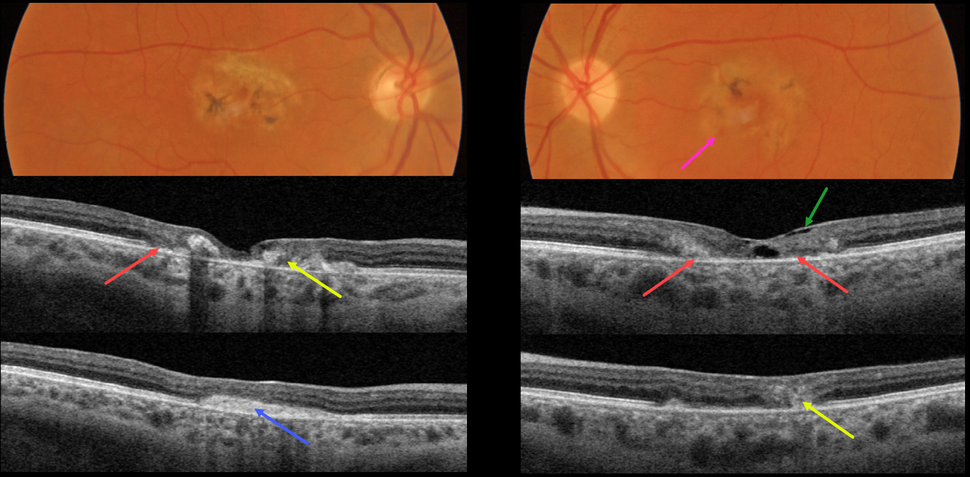
This 49YO female presented with several weeks of vision loss in her left eye. A macular neovascularization (MNV) in her right eye was treated 4 years earlier with intravitreal Avastin. Vision was 20/200 OD and 20/70 OS.
Both maculas had central pigment loss with intraretinal pigment migration. A small dot of blood was noted inferonasally in the left macula.
OCT scanning shows outer retinal atrophy, pericentral retinal disorganization, a fibrosed, inactive MNV in the right macula, and hyporeflective loss of retinal tissue (cavitation) in the left outer macula. Faint loss of the temporal inner retinal tissue is highlighted by intact internal limiting membrane (ILM drape sign).
Anti-VEGF therapy was started for a presumed MNV in her left eye.
Learning Points:
Originally described by Gass (Arch Ophthalmology 1982;100:769-780), MacTel2 is a neurodegenerative disorder, most likely originating from Muller cell dysfunction.
The term, telangiectasia, is misleading in that the funduscopic findings are mostly non-vascular. The refractile inner retinal ‘crystals’ are thought to be Muller cell footplates. The foveal retina shows a whitish discoloration, most likely due to retinal opacification caused by dysfunctional Muller cells.
Photoreceptor loss allows RPE cells to migrate along intraretinal capillaries, causing the coarse clinical pigment clumping.
All of these changes are usually in the temporal fovea, so our patient is atypical in that they involve the entire central macular regions.



