

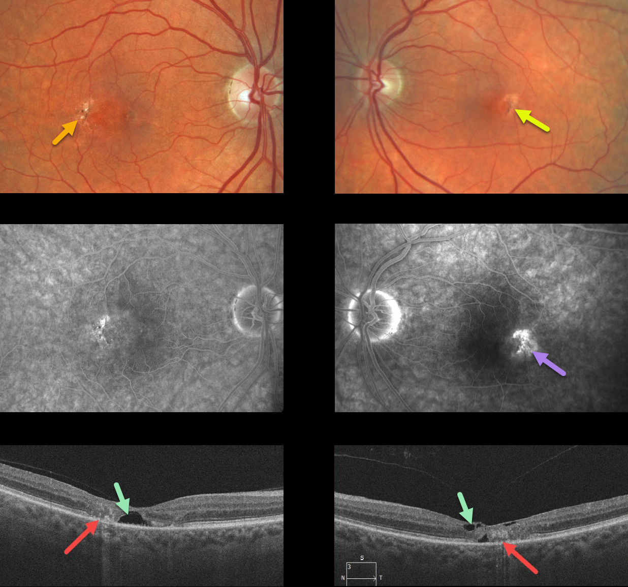
Our patient shows classic bilateral findings of macular telangiectasia type 2 (MacTel2) with temporal foveal intraretinal pigment migration and inner retinal crystals, as well as angiographic staining and leakage.
Our patient’s optical coherence tomography scans also show classic findings of mostly temporal foveal retinal atrophy, disorganization, and hyporeflective tissue loss (cavitations).
Learning Points:
MacTel type 2 is a neurodegenerative disorder, most likely originating from Müller cell dysfunction. The term telangiectasia is misleading because the funduscopic findings are mostly nonvascular.
It is thought that photoreceptor loss allows RPE cells to migrate along intraretinal capillaries, creating the pigment clumping. This is a similar mechanism to that found in other causes of intraretinal pigment migration, including photoreceptor loss or damage, such as retinitis pigmentosa, chronic retinal detachment, and blunt trauma.
The refractile inner retinal ‘crystals’ are thought to be Muller cell footplates, similar to the refractile dots found in retinoschisis.
Although much remains to be learned about its pathophysiology and there is no treatment for the underlying disease, secondary macular neovascularization can be successfully treated according to age-related macular degeneration protocols.



