

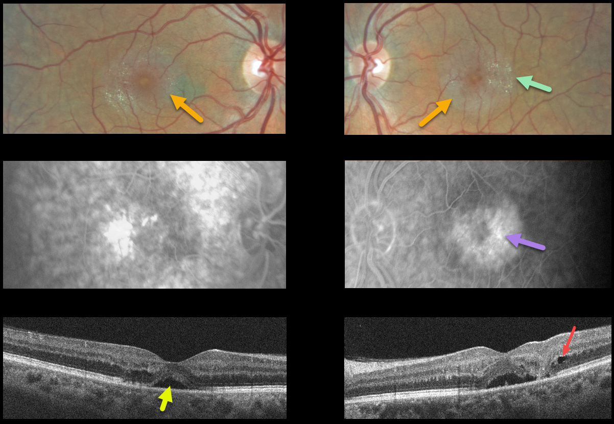
This 52YO male presented with 20/70 vision OD, 20/100 vision OS, and many classic findings for macular telangiectasia type 2 (MacTel2).
The perifoveal retina is somewhat opaque with inner retinal crystals and deep retinal angiographic leakage.
OCT scanning of the right eye shows some temporal outer retinal atrophy with an atypical small serous detachment. OCT of the left eye shows somewhat similar findings, along with a small temporal outer nuclear layer cyst vs cavitation. The serous detachment in the left fovea may represent an atypical outer macular cavitation.
Learning Points:
Originally described by Gass (Arch Ophthalmology 1982;100:769-780), MacTel2 is a neurodegenerative disorder, most likely originating from Muller cell dysfunction.
The term telangiectasia is misleading because the funduscopic findings are mostly nonvascular. The refractile inner retinal ‘crystals’ are thought to be Muller cell footplates. The foveal retina shows a whitish discoloration, most likely due to retinal opacification resulting from Müller cell dysfunction. Photoreceptor loss allows RPE cells to migrate along intraretinal capillaries, causing the coarse clinical pigment clumping (not seen in our patient).
The majority of patients will also have subclinical chorioretinal anastomosis associated with right-angle venules (Spaide et al., Retina 2018;38:1920-1929). Secondary macular neovascularization can be successfully treated according to age-related macular degeneration protocols.



