

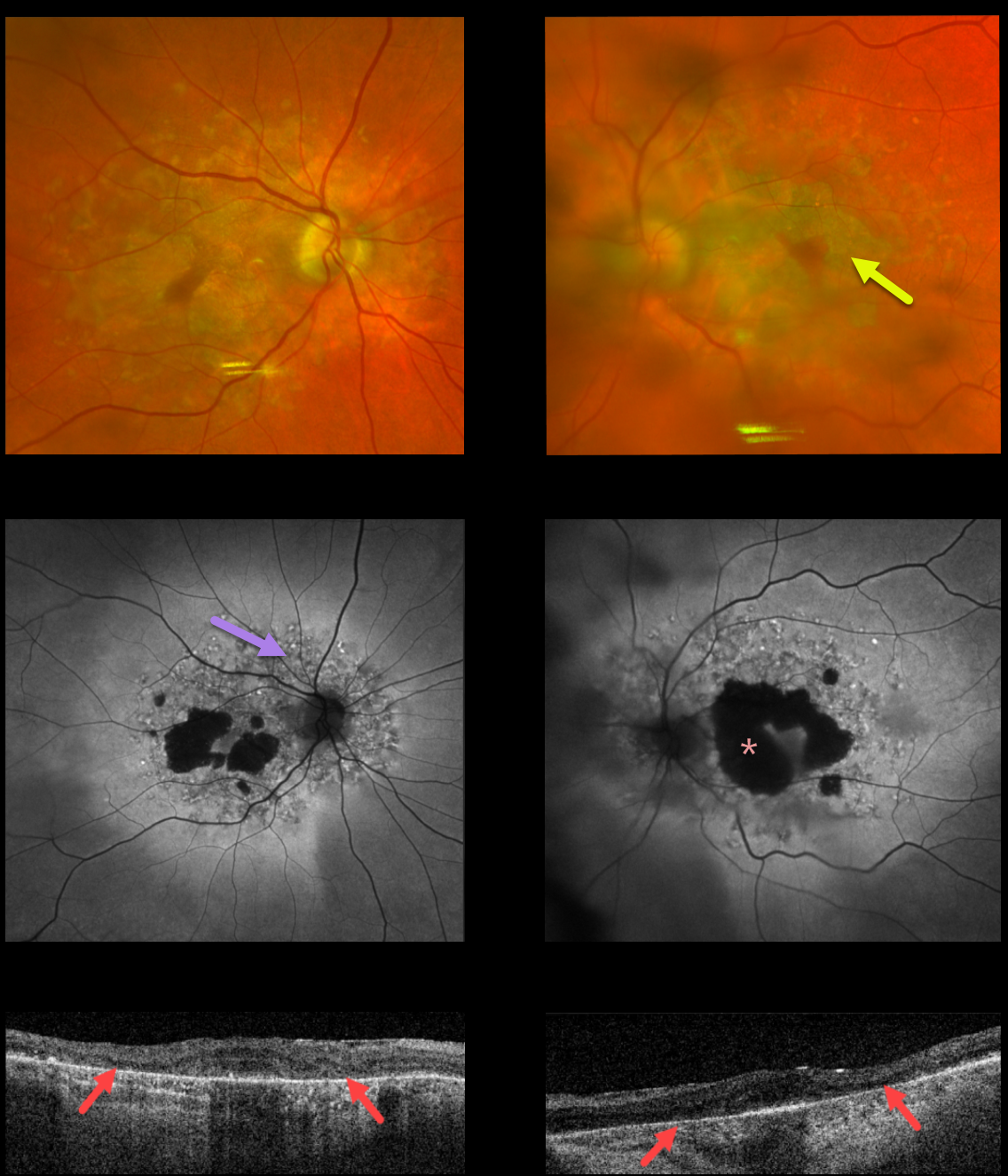
This 75YO male presented with 20/80 vision OU. Optos color imaging shows bilateral, relatively confluent patchy areas of macular atrophy.
Fundus autofluorescence (FAF) shows hypo-FAF within the areas of atrophy, with more extensive peripapillary and macular variably hyper-FAF spots.
Triton swept-source OCT shows abnormal macular architecture throughout, with the most pronounced outer retinal atrophy nasally and temporally within the areas of clinical atrophy.
Our patient has non-insulin-dependent type 2 diabetes, but there are no hearing abnormalities. MIDD patients need to be screened for other potential co-existing medical conditions, including cardiac arrhythmias, myopathy, and renal disease.
Learning Points:
Maternally inherited diabetes and deafness (MIDD) accounts for up to 3% of all cases of diabetes and results from a mutation in mitochondrial DNA at position A3243G.
MIDD often masquerades as a pattern macular dystrophy. In our experience, fundus autofluorescence (FAF) is the best way to visualize these changes. The FAF appearance somewhat resembles that seen with Elmiron toxicity.
Peripapillary hypoautofluorescence, more densely packed macular autofluorescent changes, and earlier central macular involvement suggest Elmiron toxicity over other causes (see Barnes et al., Ophthalmology Retina 2020;4:1196-1201). Note that our case spares the central macula and peripapillary retina, which is more consistent with MIDD.



