

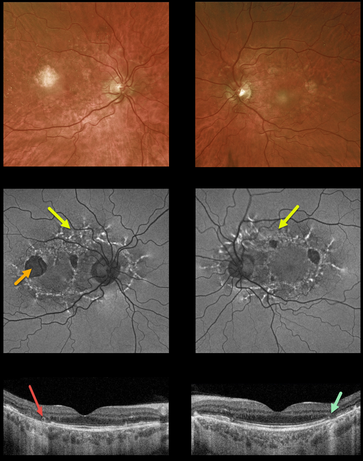
This 59YO male presented with vision of 20/400 OD and 20/30 OS. He has severe lifelong hearing loss, and both he and his mother have type 2 diabetes.
Optos ultrawidefield imaging shows temporal macular atrophy. Fundus autofluorescence shows the true extent of the pathology, with linear interconnected subretinal streaks of hyper-FAF associated with areas of hypo-FAF macular atrophy.
Swept-source OCT shows variable, mostly temporal outer retinal and RPE atrophy, along with a small area of temporal outer retinal tubulation (ORT) OD.
Learning Points: Maternally inherited diabetes and deafness (MIDD) is responsible for up to 3% of all cases of diabetes, and results from a mutation of mitochondrial DNA A3243G. MIDD often masquerades as a pattern macular dystrophy.
Fundus autofluorescence (FAF) in our experience is the best way to visualize these changes. The FAF appearance somewhat resembles that seen with Elmiron toxicity.
Peripapillary hypoautofluorescence, more densely-packed macular autofluorescent changes, and earlier central macular involvement suggest Elmiron toxicity over other causes (see Barnes et al Ophthalmology Retina 2020;4:1196-1201). Note that our case spares the central macula and peripapillary retina, which is more consistent with MIDD.
ORT is often noted overlying inactive MNV with ongoing anti-VEGF therapy and should not be confused with exudative fluid or cysts which lack a hyperreflective border. The outer hyperreflective band likely represents inner segment mitochondria undergoing fission and translocation toward the nucleus (Litts et al, Retina 2018;38:445-461).
ORT, initially described by Zweifel et al (Arch Ophthalmol 2009;127:1596-1602), is a neurodegenerative condition of the photoreceptors and Muller cells associated with atrophy affecting the outer retina and retinal pigment epithelium, including advanced AMD and inherited retinal diseases.



