

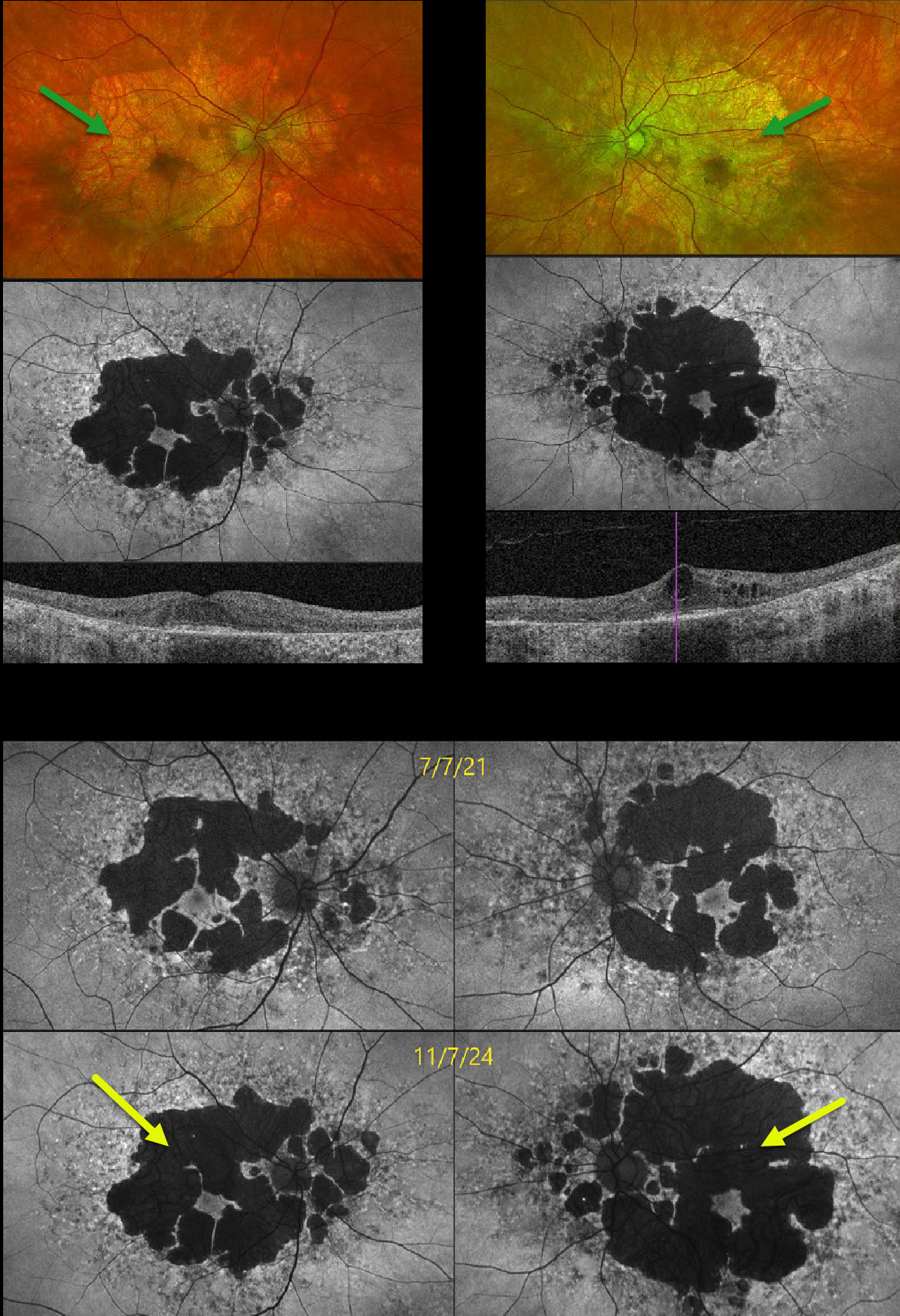
This 58YO male presented with several years of progressive vision loss. He has a history of well-controlled type 2 diabetes. Vision was 20/30 OD and 20/60 OS.
Optos color RG imaging shows fairly symmetric areas of variable, confluent, non-central macular atrophy. Fundus autofluorescence (FAF) shows hypo-FAF from the areas of atrophy, along with areas of hyper-FAF around each nerve and macula. Swept-source OCT shows outer retinal and RPE atrophy bilaterally with central cystic edema OS. FAF shows significant progression of atrophy compared with 3 years earlier. The subclinical edema OS was felt to be diabetic and has been observed since it was spontaneously fluctuating with unchanged vision and symptoms.
Learning Points:
Maternally inherited diabetes and deafness (MIDD) accounts for up to 3% of all cases of diabetes and results from the A3243 G mutation in mitochondrial DNA. MIDD often masquerades as a pattern macular dystrophy. In our experience, fundus autofluorescence (FAF) is the best way to visualize these changes. The FAF appearance somewhat resembles that seen with Elmiron toxicity. Peripapillary hypoautofluorescence, more densely packed macular autofluorescent changes, and earlier central macular involvement suggest Elmiron toxicity over other causes (Barnes et al Ophthalmology Retina 2020;4:1196-1201).
Despite our patient’s multimodal findings being classical for MIDD, mitochondrial genetic testing revealed variants of uncertain significance (homozygous AMPD1 and heterozygous ACAD5). Invitae Inherited Retinal Disorders Panel revealed homozygous variants of u



