

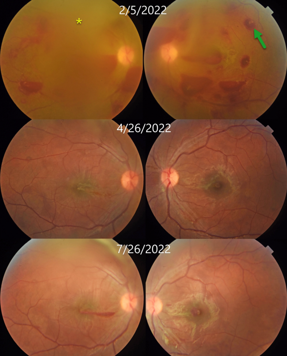
This 17YO male presented with 1 week of bilateral vision loss. Vision was counting fingers OU.
Bilateral white-centered and preretinal hemorrhages are noted. The fundus detail is decreased bilaterally due to breakthrough vitreous blood. Bloodwork revealed severe anemia with a hemoglobin of 5.3 g/dl. Vitamin B12 level was decreased at 160 pg/ml (normal 187-833). Based on the peripheral blood smear, which showed macrocytes, anisocytosis, poikilocytosis, target cells, and nucleated red blood cells, he was diagnosed with megaloblastic anemia.
His B12 deficiency resulted from a poorly balanced vegetarian diet. Following several blood transfusions and injectable vitamin B12 supplementation, as well as oral sublingual B12 and dietary modification, his anemia improved.
Two months later, vision improved to 20/200 OD and 20/80 OS; the hemoglobin rose to 12 g/dL; and funduscopic findings were improving. Five months following presentation, vision improved to 20/40 OU, and the retinal findings continued to normalize.
Learning Points:
Megaloblastic anemia is characterized by low hemoglobin in the presence of megaloblasts (large and immature nucleated red blood cells). It is caused by B12 or B9 (folate) deficiency, either from poor dietary intake (as in our patient) or from structural/functional gastrointestinal disorders that impair vitamin B malabsorption.
White-centered retinal hemorrhages and sub-internal limiting membrane/preretinal blood are commonly associated with anemia and blood dyscrasias. Other causes include bacterial endocarditis, hypertension, diabetic retinopathy, HIV retinopathy, and shaken baby syndrome. Hence, comorbidity must be questioned in such cases, and the patient should be screened for systemic diseases.



