

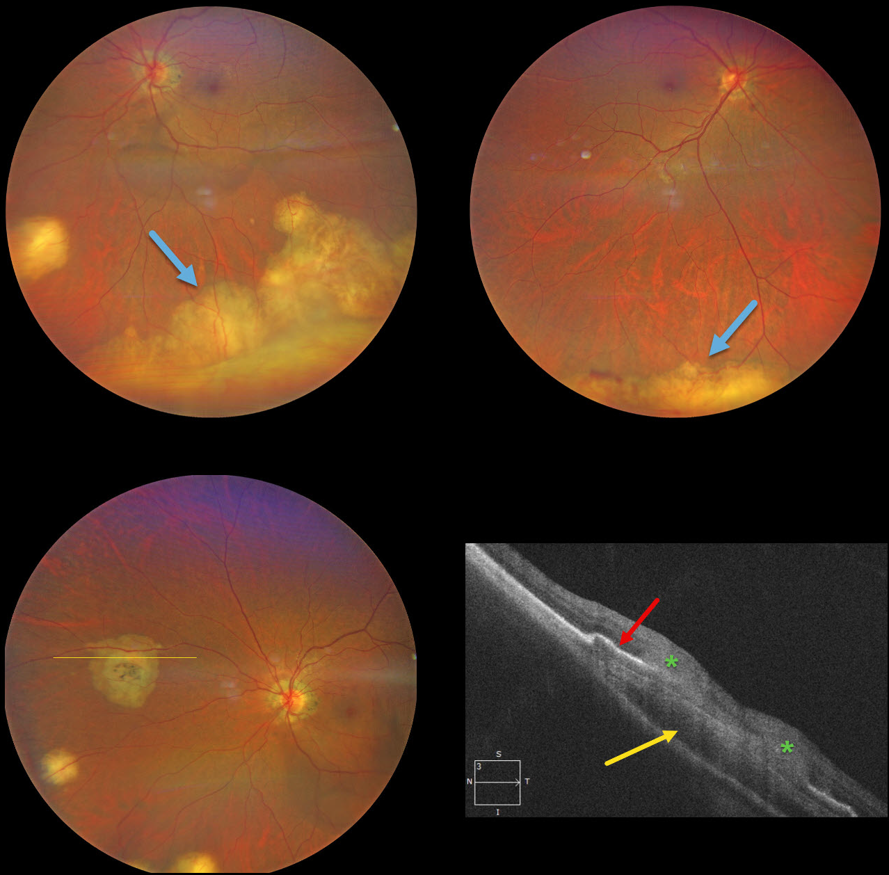
This previously healthy 49YO Indian female was visiting the United States and presented with new floaters in her left eye. Vision was 20/25 OD and 20/40 OS. There was no anterior uveitis, and the vitreous was clear bilaterally.
Color photography shows multifocal bilateral amelanotic choroidal lesions, some isolated and others confluent in the inferior periphery. OCT scanning through a nasal lesion OS shows a relatively hyporeflective choroidal lesion, irregular overlying RPE elevation, and probable infiltration of the overlying retina.
QuantiFERON-TB testing was positive with a negative chest X-ray. The patient was referred to the infectious disease department and started on anti-tuberculosis treatment (ATT).
Learning Points:
Although TB most commonly presents with pulmonary involvement, extrapulmonary sites can include the gastrointestinal, skin, cardiovascular, genitourinary, and central nervous systems, as well as the eyes. Ocular involvement, like syphilis, can mimic virtually any type of uveitis, including anterior, intermediate, posterior, and panuveitis, retinitis and retinal vasculitis, neuroretinitis, optic neuropathy, choroidal granuloma (our patient), choroiditis, and scleritis. For an excellent review of intraocular tuberculosis, see Bupta et al, Survey Ophthalmology 2007;52:561-587.
Tubercular granulomas can be distinguished from sarcoid lesions by being solitary, yellow, larger, and vascularized (Agarwal et al, AJO 2021;226:42-55). Our patient is therefore somewhat unusual with bilateral multifocal lesions. Sarcoid workup was negative.



