

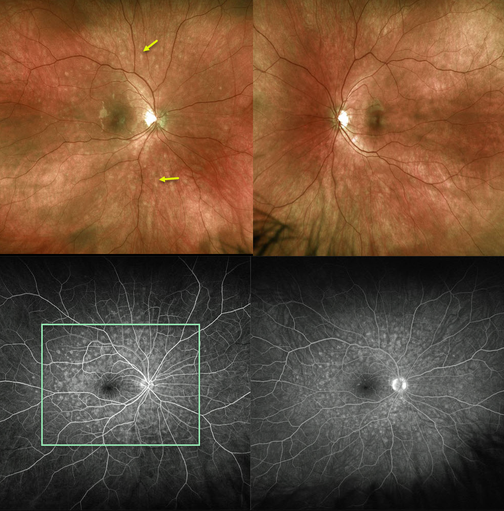
The Optos fundus photo of this 20YO female shows 100-200 micron outer retinal white spots scattered around the posterior pole, characteristic of multiple evanescent white dot syndrome (MEWDS).
Fluorescein angiography shows a wreath-like pattern of hyperfluorescence.
Learning Points:
MEWDS is an idiopathic, usually unilateral inflammation of the outer retina.
Most common in young women, patients often have a preceding viral syndrome and complain of blurred vision, photopsias, and temporal visual field loss.
Initially presenting with scatttered outer retinal white spots, the spots resolve within a few weeks making the diagnosis elusive. However, the enlarged blind spot often remains for months.
The condition is self-limited and usually resolves within several months without treatment.



