

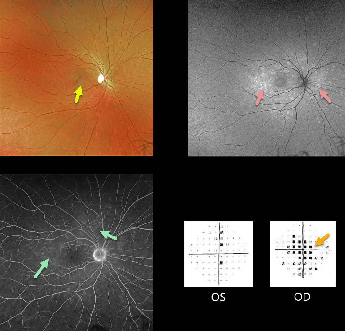
Our patient is a 31YO female with acute vision loss of 20/60 in her right eye with classic MEWDS findings.
Granular foveal changes (Jampol spots) and scattered white spots are seen in the outer retina. The spots appear hyper-autofluorescent on fundus autofluorescence imaging.
Fluorescein angiography shows multifocal wreath-like hyperfluorescent staining lesions.
Acute cases will also have an enlarged blind spot, often with temporal visual field loss.
Learning Points:
The MEWDS white dots usually disappear within a week or two, which is why the lesions are called evanescent. However, the blind spot may persist for many months, well after the fundus appearance has normalized.
This stage of MEWDS was originally called the Acute idiopathic blind spot enlargement (AIBSE) syndrome before Gass recognized that the 2 disorders were the same disease but with distinct findings at different points in time.



