

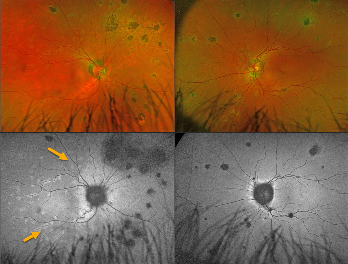
This 56YO female presented with 1 week of flashes and blurred vision in her right eye. Vision was 20/25 OD and 20/20 OS.
Coarse, large pigmented multifocal scars with peripapillary scarring are noted OU. Multiple deep white retinal spots are scattered throughout the right posterior pole. These spots on fundus autofluorescence (FAF) hyper-autofluoresce. Confluent hyper-FAF extends outwards from the optic nerve.
Learning Points:
Multiple evanescent white dot syndrome (MEWDS) is a usually unilateral, idiopathic inflammation of the outer retina that typically occurs in young, healthy myopic females. Acutely, patients will also have an enlarged blind spot, often complaining of temporal visual field loss, due to the peripapillary inflammation. The white dots disappear within a week or two, which is why the lesions are called “evanescent”.
Multifocal choroiditis (MFC) is the current preferred term for a group of historically heterogeneous disorders, including multifocal choroiditis, recurrent multifocal choroiditis, multifocal choroiditis and panuveitis, punctate inner choroidopathy, progressive subretinal fibrosis, and pseudo-histoplasmosis (Essex et al, Retina 2013;33:1-4 and Spaide et al, Retina 2013;33:1315-1324). MFC occurs in otherwise healthy adults, often myopic and female. Findings include variably pigmented multifocal scars throughout the fundus, panuveitis, subretinal fibrosis, and macular neovascularization (MNV). Unlike ocular histoplasmosis, new scars and uveitis can be seen. MNV can be successfully managed with anti-VEGF therapy.
Our patient’s scars represent either preexisting MFC or ocular histoplasmosis, and MEWDS is sometimes found in some of these eyes. Distinct from primary MEWDS, secondary MEWDS may be an epiphenomenon in inflammatory conditions such as MFC that disrupt the choriocapillaris-Bruch membrane-RPE complex (Essilfie et al, Retina 2022;42:227-235).



