

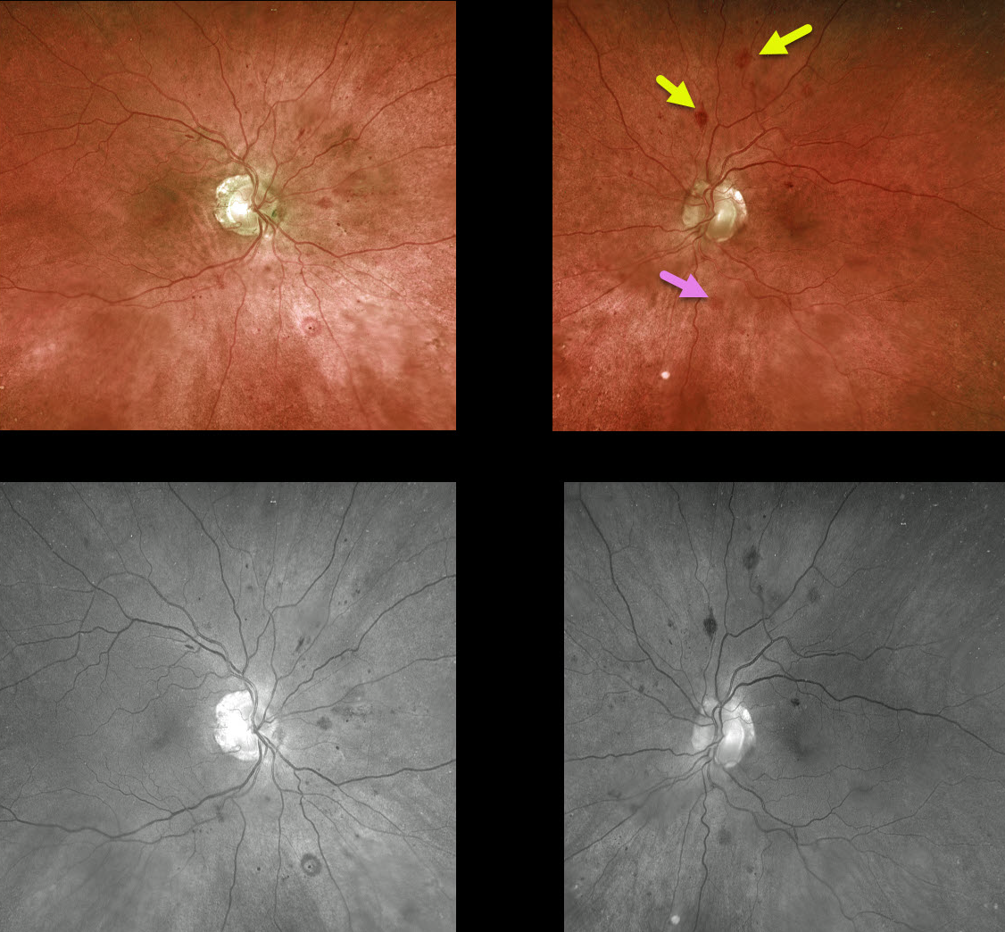
This 76YO male presented with normal vision and asymptomatic bilateral retinal hemorrhages following a recent diagnosis of multiple myeloma.
There are a variety of retinal hemorrhages seen on Optos and green-channel images, including inner retinal white-centered hemorrhages and deep blot hemorrhages in the outer plexiform layer.
Learning Points:
White-centered hemorrhages or “roth spots” can be seen in many disorders, including leukemia, hyperviscosity, anemia, diabetes, and hypertension. The white material may represent a variety of causes, including fibrin.
The term “Roth spots” should thus be reserved only for patients with bacterial endocarditis. See Duane et al Ophthalmology 1980;87;66-69.
The deeper hemorrhages would be unusual for the more common disorders seen in clinical practice (including diabetic retinopathy, retinal vein occlusions, and hypertensive retinopathy), and are usually indicative of blood dyscrasias, including anemia and hematologic malignancies. These deeper hemorrhages are also seen peripherally in ocular ischemia.



