

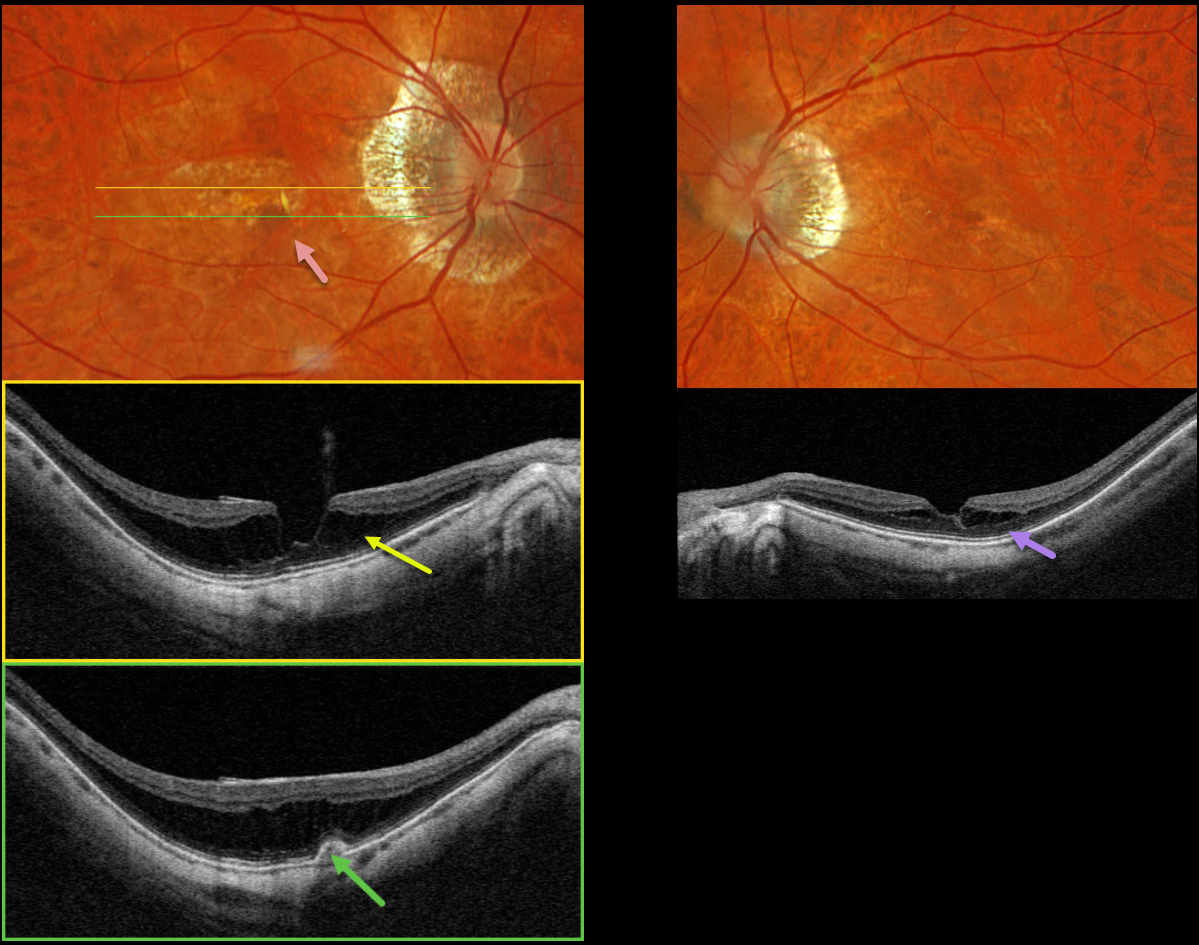
This 72YO female recently moved to our area and self-referred herself to establish new retinal care. One year earlier, she received a series of five monthly anti-VEGF injections OD for myopic macular neovascularization (MNV), and felt that her vision was stable since the last injection about 6 months earlier. Vision was 20/60 OD and 20/40 OS.
Optos color RGB imaging OD shows myopic peripapillary atrophy, central macular pigment loss, and a pigmented subretinal lesion.
Triton swept-source OCT through the macular center shows a shallow staphylomatous posterior bowing of the sclera with overlying outer macular schisis. The choroid is diffusely thinned. Scanning the pigmented lesion reveals a small type 1 MNV (below the RPE) without fluid. Imaging of her left eye shows much milder, central schisis in the outer plexiform layer. Observation was recommended.
Learning Points:
In our experience, myopic MNV presents quite differently from those with neovascular AMD. Significant subretinal blood, subretinal fluid or intraretinal fluid are rarely seen.
Often, an active MNV will present as a new dot of subretinal blood or pigment. Occasionally, there will just be symptomatic vision loss or metamorphopsia without clinical or OCT findings.
Anti-VEGF therapy is effective, and these lesions can often be managed with PRN therapy (Sakata et al, Retina 2023;43:1863-1871) as in our case.



